Search results (32 results)
-
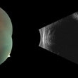
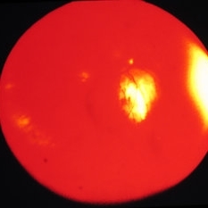
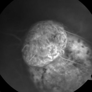 Collar Button Ocular Melanoma-FA
Feb 22 2015 by Jeffrey G. Gross, MD, FASRS
68-year-old male with ocular melanoma associated with exudative retinal detachment. Vision 20/100.
Photographer: Tammy Pittman
Imaging device: Visucam
Condition/keywords: exudative retinal detachment, melanoma
-
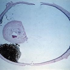
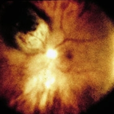
 Collar Button Choroidal Melanoma
Oct 25 2015 by Dwain G. Fuller, MD, JD
Ultrasonographic image of collar button choroidal melanoma with associated serous retinal detachment.
Photographer: Jana Sierocki
Condition/keywords: collar button, malignant melanoma
-
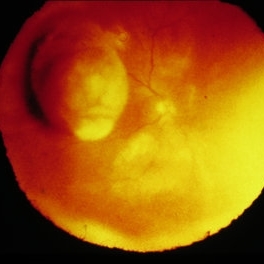
 Choroidal Melanoma
Sep 6 2013 by Theodore Leng, MD, MS, FASRS
30-year-old male with a collar button mushroom shaped choroidal melanoma and associated exudative retinal detachment.
-
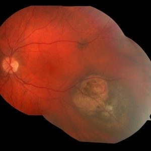
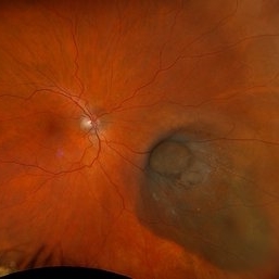
 Collar Button Choroidal Melanoma
Oct 25 2015 by Dwain G. Fuller, MD, JD
Fundus photograph of collar button choroidal melanoma with associated serous retinal detachment and pre-retinal hemorrhages.
Condition/keywords: collar button, malignant melanoma
-
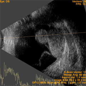 Collar Button Appearance on B-Scan
Aug 28 2019 by Gayathri Mohan
B-scan showing an intraocular mass with collar button appearance. Suspected case of choroidal melanoma.
Photographer: Dr.Gayathri Mohan, Retina Foundation
Imaging device: Nidek Mirante SLO
Condition/keywords: choroidal mass, collar button
-
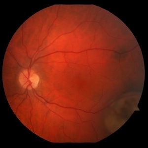
 Collar Button Choroidal Melanoma
Oct 25 2015 by Dwain G. Fuller, MD, JD
Fundus photograph of collar button choroidal melanoma.
Imaging device: Optos
Condition/keywords: collar button, malignant melanoma
-
 Wide Field Fundus Montage of Intraocular Mass with Retinal Detachment
Aug 28 2019 by Gayathri Mohan
50 year old female came with diminution of vision in the LE. Wide field fundus photograph showing an intraocular mass temporally along with an exudative retinal detachment inferiorly. Ultrasonography showed an intraocular mass with collar button appearance suggestive of a Choroidal melanoma. She underwent enucleation and histopathology confirmed a spindle cell choroidal melanoma
Photographer: Dr. Gayathri Mohan, Retina Foundation
Imaging device: Nidek Mirante SLO
Condition/keywords: choroidal mass, collar button
-
 Melanoma
Feb 28 2013 by Theodore Leng, MD, MS, FASRS
30-year-old male with a collar button mushroom shaped choroidal melanoma and associated exudative retinal detachment.
Imaging device: Zeiss FF450
-
 Choroidal Melanoma
Sep 6 2013 by Theodore Leng, MD, MS, FASRS
30-year-old male with a collar button mushroom shaped choroidal melanoma and associated exudative retinal detachment.
-
Choroidal Melanoma
Sep 6 2013 by Theodore Leng, MD, MS, FASRS
30-year-old male with a collar button mushroom shaped choroidal melanoma and associated exudative retinal detachment.
-
 B-Scan Showing Intraocular Mass
Aug 28 2019 by Gayathri Mohan
50 year old female came with diminution of vision in the LE. Ultrasonography showed an intraocular mass with collar button appearance suggestive of a Choroidal melanoma. She underwent enucleation and histopathology confirmed a spindle cell choroidal melanoma
Photographer: Dr. Gayathri Mohan - Retina Foundation
Imaging device: Nidek ,Mirante
Condition/keywords: collar button, melanoma
-
 Collar Button Choroidal Melanoma
Oct 25 2015 by Dwain G. Fuller, MD, JD
Fundus photograph of large collar button choroidal melanoma.
Condition/keywords: collar button, malignant melanoma
-
 Juxtapapillary Choroidal Melanoma
Oct 25 2015 by Dwain G. Fuller, MD, JD
Fundus photograph and b-scan ultrasonographic image of a collar button choroidal melanoma with prominent intrinsic vascularity.
Photographer: Ultrasound: Jana Sierocki
-
 Collar Button Melanoma
Mar 27 2025 by Virginia Gebhart
62 year old male with large pigmented lesion with collar button. Pt states he was never aware of any lesion/nevus in the past. Fluid and orange pigment present, appears to be chronic. Pt will be scheduled for brachytherapy pending CT scan results.
Photographer: Virginia Gebhart, Retina Consultants of Carolina
Imaging device: Optos California
Condition/keywords: choroidal melanoma, collar button
-
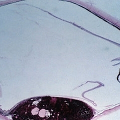
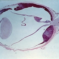
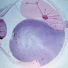
 Slide 14-13
Mar 4 2019 by Lancaster Course in Ophthalmology
Among the objective manifestations of a choroidal melanoma, the most important is the presence of a solid mass seen on ophthaloscopic presentation. (seen in Slides 14-1 to 14-13). The shape and projection of the tumor can be best appreciated by observation with the indirect ophthalmoscope. The appearance of these tumors is typically that of a mushroom-shaped, collar button-shaped mass.
Condition/keywords: melanoma, tumor
-
 Slide 14-8
Mar 14 2019 by Lancaster Course in Ophthalmology
Among the objective manifestations of a choroidal melanoma, the most important is the presence of a solid mass seen on ophthaloscopic presentation. (seen in Slides 14-1 to 14-13). The shape and projection of the tumor can be best appreciated by observation with the indirect ophthalmoscope. The appearance of these tumors is typically that of a mushroom-shaped, collar button-shaped mass.
Condition/keywords: melanoma, tumor
-
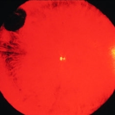
 Choroidal Melanoma
Oct 27 2023 by Virginia Gebhart
76 year old male with suspicious pigmented choroidal lesion with new collar button growth. Blocking defect and vascularity noted on FA
Photographer: Virginia Gebhart
Condition/keywords: FA late phase, fluorescein angiogram (FA), Fluorescein angiography, melanoma
-
 Slide 14-10
Mar 4 2019 by Lancaster Course in Ophthalmology
Among the objective manifestations of a choroidal melanoma, the most important is the presence of a solid mass seen on ophthaloscopic presentation. (seen in Slides 14-1 to 14-13). The shape and projection of the tumor can be best appreciated by observation with the indirect ophthalmoscope. The appearance of these tumors is typically that of a mushroom-shaped, collar button-shaped mass.
Condition/keywords: melanoma, tumor
-
 Choroidal Melanoma
Mar 1 2024 by Virginia Gebhart
52 year old female at first visit July 2023 vs 7 months s/p brachytherapy. SRF in macula has resolved, trace fluid on posterior edge of collapsing collar button.
Photographer: Virginia Gebhart
Imaging device: Optos California
Condition/keywords: brachytherapy, Choroidal melanoma, collar button
-
 Slide 14-12
Mar 4 2019 by Lancaster Course in Ophthalmology
Among the objective manifestations of a choroidal melanoma, the most important is the presence of a solid mass seen on ophthaloscopic presentation. (seen in Slides 14-1 to 14-13). The shape and projection of the tumor can be best appreciated by observation with the indirect ophthalmoscope. The appearance of these tumors is typically that of a mushroom-shaped, collar button-shaped mass.
Condition/keywords: melanoma, tumor
-
 Slide 14-3
Mar 4 2019 by Lancaster Course in Ophthalmology
Among the objective manifestations of a choroidal melanoma, the most important is the presence of a solid mass seen on ophthaloscopic presentation. (seen in Slides 14-1 to 14-13). The shape and projection of the tumor can be best appreciated by observation with the indirect ophthalmoscope. The appearance of these tumors is typically that of a mushroom-shaped, collar button-shaped mass.
Condition/keywords: melanoma, tumor
-
 Slide 14-7
Mar 4 2019 by Lancaster Course in Ophthalmology
Among the objective manifestations of a choroidal melanoma, the most important is the presence of a solid mass seen on ophthaloscopic presentation. (seen in Slides 14-1 to 14-13). The shape and projection of the tumor can be best appreciated by observation with the indirect ophthalmoscope. The appearance of these tumors is typically that of a mushroom-shaped, collar button-shaped mass.
Condition/keywords: melanoma, tumor
-
 Slide 14-9
Mar 4 2019 by Lancaster Course in Ophthalmology
Among the objective manifestations of a choroidal melanoma, the most important is the presence of a solid mass seen on ophthaloscopic presentation. (seen in Slides 14-1 to 14-13). The shape and projection of the tumor can be best appreciated by observation with the indirect ophthalmoscope. The appearance of these tumors is typically that of a mushroom-shaped, collar button-shaped mass.
Condition/keywords: melanoma, tumor
-
 Slide 14-5
Mar 4 2019 by Lancaster Course in Ophthalmology
Among the objective manifestations of a choroidal melanoma, the most important is the presence of a solid mass seen on ophthaloscopic presentation. (seen in Slides 14-1 to 14-13). The shape and projection of the tumor can be best appreciated by observation with the indirect ophthalmoscope. The appearance of these tumors is typically that of a mushroom-shaped, collar button-shaped mass.
Condition/keywords: melanoma, tumor
-
 Slide 14-1
Mar 4 2019 by Lancaster Course in Ophthalmology
Among the objective manifestations of a choroidal melanoma, the most important is the presence of a solid mass seen on ophthaloscopic presentation. (seen in Slides 14-1 to 14-13). The shape and projection of the tumor can be best appreciated by observation with the indirect ophthalmoscope. The appearance of these tumors is typically that of a mushroom-shaped, collar button-shaped mass.
Condition/keywords: melanoma, tumor
 Loading…