Search results (34 results)
-
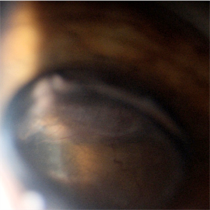
 Ciliary Body Melanoma With Partial Ring Configuration and Diffuse Sentinel Vessels
Feb 26 2014 by Susanna S. Park, MD, PhD
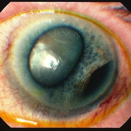
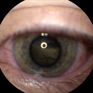
Slit lamp photo of a 57-year-old man with new vision loss from cataract formation. Large ciliary body mass with diffuse sentinel vessels is noted. The eye was removed and the tumor was noted to have a partial ring configuration with predominantly epithelioid cells and early vitreous seeding.
Photographer: Ellen Redenbo, University of California Davis Eye Center
Condition/keywords: ciliary body melanoma, melanoma
-
 Ciliary Body Melanoma B-Scan Ultrasound
Feb 27 2014 by Susanna S. Park, MD, PhD
Large ciliary body melanoma in a 57-year-old man.
Photographer: Ellen Redenbo, University of California Davis Eye Center
Condition/keywords: B scan ultrasound, ciliary body melanoma
-
 Ciliary Body Melanoma
Jul 12 2013 by Jason S. Calhoun
71-year-old male who was recently diagnosed with a large ciliary body melanoma that is pushing into the anterior chamber of the left eye. Patient is going to proceed with proton therapy.
Photographer: Jason S. Calhoun, Department of Ophthalmology, Mayo Clinic Jacksonville, Florida
Condition/keywords: ciliary body melanoma
-
 Ciliary Body Ocular Melanoma
May 9 2016 by Nichole Lewis
Ciliary body ocular melanoma.
Photographer: Nichole Lewis
Condition/keywords: ciliary body melanoma
-
 Ciliary Body Melanoma-UBM
Feb 27 2014 by Susanna S. Park, MD, PhD
UBM of a large ciliary body melanoma in a 57-year-old man. Histopathology after eye removal showed a diffuse ring component that also involved the anterior choroid.
Photographer: Ellen Redenbo, University of California Davis Eye Center
Condition/keywords: ciliary body melanoma, ultrasound
-
 Ciliary body melanoma
Jan 11 2013 by Alex P. Hunyor, MD
Left inferotemporal ciliary body melanoma with displacement of pupil, cataract, and large dilated episcleral vessels.
-
 Ciliary body melanoma
May 2 2013 by Henry J. Kaplan, MD
Ciliary body melanoma visible through the pupil.
Condition/keywords: ciliary body melanoma
-
 Ciliary Body Melanoma
Jul 12 2013 by Jason S. Calhoun
71-year-old male who was recently diagnosed with a large ciliary body melanoma that is pushing into the anterior chamber of the left eye. Patient is going to proceed with proton therapy.
Photographer: Jason S. Calhoun, Department of Ophthalmology, Mayo Clinic Jacksonville, Florida
Condition/keywords: ciliary body melanoma
-
 Sentinel Vessel in Ciliary Body Melanoma
Apr 1 2019 by Gary R. Cook, MD, FACS
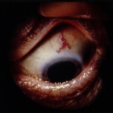
White male with a sentinel vessel OD due to the presence of a ciliary body melanoma; VA = 20/30-2.
Imaging device: Topcon VT-50
Condition/keywords: ciliary, melanocytic lesion, melanoma, sentinel vessel
-
 Ciliary Body Choroidal Melanoma
Jan 29 2015 by H. Michael Lambert, MD
Invivo presentation of ciliary body melanoma.
Condition/keywords: ciliary body melanoma
-
 Ciliary Body Choroidal Melanoma
Jan 29 2015 by H. Michael Lambert, MD
Large mass hanging posterior to the lens superiorly in a dilated eye.
Condition/keywords: ciliary body melanoma
-
 Choroidal and Ciliary Body Melanoma
Apr 11 2018 by Jason Griffith
15-year-old male patient referred for suspicious mass.
Photographer: Jason Griffith, Tennessee Retina, Nashville, TN
Imaging device: Topcon TRC 50EX
Condition/keywords: ciliary body melanoma
-
 Ciliary Body Melanoma
Apr 1 2019 by Gary R. Cook, MD, FACS
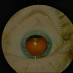
White male with a ciliary body melanoma OD seen as a dark, dome-shaped mass through a dilated pupil; VA=20/30-2.
Imaging device: Topcon VT-50
Condition/keywords: ciliary body mass, melanocytic lesion, melanoma
-
 Ciliary Body Melanoma (Class 2 PRAME Positive)
Jan 17 2019 by Olivia Rainey
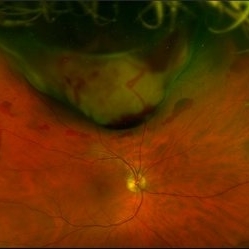
Pseudocolor optos fundus image of an 61-year-old male with ciliary body melanoma affecting his right eye, which tested PRAME positive placing him in class 2. The melanoma had caused a hemorrhagic retinal detachment. Patient was referred for a retinal detachment and was experiencing cloudy vision affecting his inferior field. He had been seeing flashes inferiorly 1-2x a daily for about 6-8 months.
Photographer: Olivia Rainey
Imaging device: Optos
Condition/keywords: ciliary body melanoma, class 2, Optos, PRAME positive
-
 Ciliochoroidal Melanoma
May 14 2020 by Anfisa Ayalon, MD
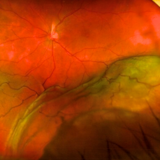
Fundus photograph of a 71-year-old woman with ciliochoroidal melanoma. Note a melanoma-associated massive exudative retinal detachment.
Photographer: Anfisa Ayalon, MD., Meir Medical Center, Kfar Saba, Israel.
Imaging device: California, Optos 200 DTX
Condition/keywords: ciliary body melanoma, exudative retinal detachment, melanoma
-
 Superior Ciliochoroidal Melanoma with Diffuse Vitreous Seeding
Jan 21 2020 by Sophia El Hamichi, MD
Montage photograph of a fundus of a superior ciliochoroidal melanoma with diffuse pigmentary vitreous seeding.
Condition/keywords: ciliary body melanoma, melanoma, montage, vitreous seeding
-
 Ciliary Body Melanoma
Jul 4 2021 by Gerardo Rivera Arroyo
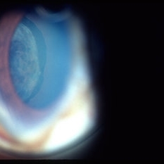
Clinical image taken in a slit lamp with a gonioscopy of a 39-year-old female patient with ciliary body melanoma before enucleation and pathological study.
Condition/keywords: ciliary body melanoma, gonioscopy
-
 Ciliary body melanoma
Jul 4 2021 by Gerardo Rivera Arroyo
Clinical image taken in a slit lamp with a gonioscopy of a 39-year-old female patient with ciliary body melanoma before enucleation and pathological study.
Condition/keywords: ciliary body melanoma, gonioscopy
-
 Fundus Autofluorescence in Ciliochoroidal Melanoma
May 14 2020 by Anfisa Ayalon, MD
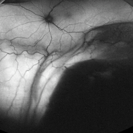
Fundus autofluorescence photograph of a 71-year-old woman with ciliochoroidal melanoma. Note a melanoma-associated massive exudative retinal detachment.
Photographer: Anfisa Ayalon, MD., Meir Medical Center, Kfar Saba, Israel.
Imaging device: California, Optos 200 DTX
Condition/keywords: ciliary body melanoma, exudative retinal detachment
-
 Ciliochoroidal Melanoma Photograph with Gonioscopy Lens
May 14 2020 by Anfisa Ayalon, MD
Gonioscopy photograph of a 71-year-old woman with ciliochoroidal melanoma. Note a melanoma-associated exudative retinal detachment and feeding vessels.
Photographer: Anfisa Ayalon, MD., Meir Medical Center, Kfar Saba, Israel.
Condition/keywords: ciliary body melanoma, exudative retinal detachment, gonioscopy
 Loading…