Search results (404 results)
-
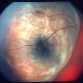
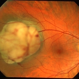 Choroidal melanoma case 4 - partly amelanotic
Jan 11 2013 by Alex P. Hunyor, MD
Choroidal melanoma with amelanotic "collar stud."
Condition/keywords: melanoma
-
 Choroidal Melanoma
Jul 4 2012 by John T. Thompson, MD
Amelanotic choroidal melanoma with serous retinal detachment
Condition/keywords: choroidal tumor, exudative retinal detachment, melanoma
-
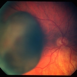
 Choroidal Melanoma Large Amelanotic
Oct 15 2012 by Susanna S. Park, MD, PhD
68-year-old man with a large amelanotic mass and subretinal fluid in the left eye. Visual acuity was CF and extrascleral extension was suspected on MRI scan.
Photographer: Ellen Redenbo, University of California Davis Eye Center
Condition/keywords: melanoma
-
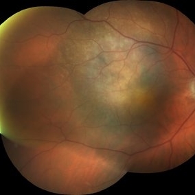
 Melanocytoma with Choroidal Melanoma
Oct 8 2012 by Susanna S. Park, MD, PhD
Fundus photograph of a 75-year-old woman with a slowly growing pigmented lesion.
Photographer: Ellen Redenbo, University of California Davis Eye Center
Condition/keywords: melanocytoma
-
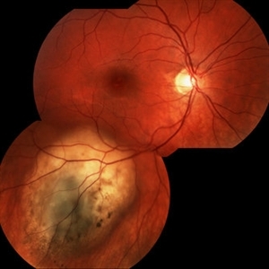
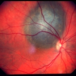 Choroidal melanoma case 3 - peripapillary
Jan 11 2013 by Alex P. Hunyor, MD
Right peripapillary choroidal melanoma.
-
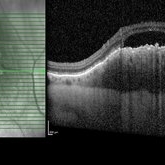
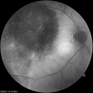
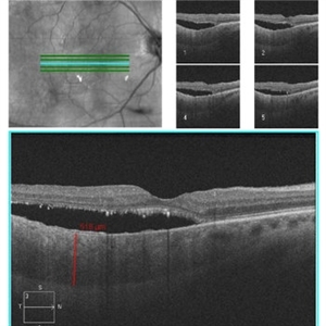 Diffuse Choroidal Melanoma OCT
Aug 24 2012 by John S. King, MD
Photographer: Kristin Konecki, OcuSight Eye Care Center, Rochester, NY
-
 Choroidal Melanoma
May 2 2013 by Henry J. Kaplan, MD
Peripapillary choroidal melanoma.
-
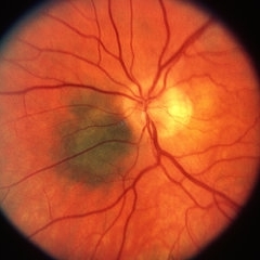
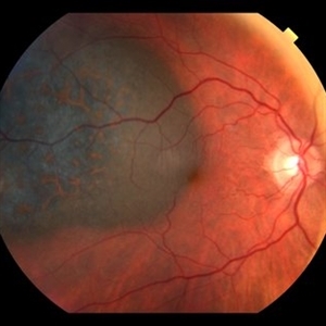
 Choroidal Melanoma
Sep 5 2012 by Virgilio Morales-Canton, MD
Fundus photograph of a 62-year-old male with a choroidal melanoma on the posterior pole. Double circulation is observed.
Photographer: Virgilio Morales-Canton
-
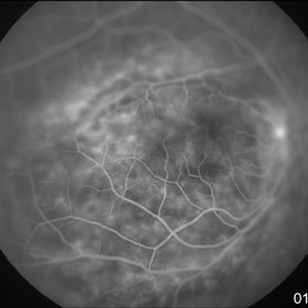
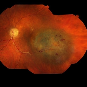
 Choroidal melanoma 1 - colour photo
Jan 11 2013 by Alex P. Hunyor, MD
Large choroidal melanoma with "collar stud" appearance. See accompanying FA showing tumour circulation.
Condition/keywords: melanoma
-
 Choroidal Melanoma Enlarged
Oct 16 2012 by Jeffrey G. Gross, MD, FASRS
Choroidal melanoma enlarged, and more pigmented, 3 months later.
-
 Choroidal melanoma color
-
 Ciliochoroidal melanoma case 2
Jan 11 2013 by Alex P. Hunyor, MD
Large right ciliochoroidal melanoma with episcleral "sentinel" vessel.
Condition/keywords: melanoma
-
 Diffuse Choroidal Melanoma
Aug 24 2012 by John S. King, MD
Diffuse Choroidal Melanoma
Photographer: Kristin Konecki, OcuSight Eye Care Center, Rochester, NY
-
 Choroidal Melanoma with Minimal Pigment
Sep 17 2012 by Susanna S. Park, MD, PhD
32-year-old otherwise healthy woman with no visual complaint.
Photographer: Ellen Redenbo, University of California Davis Eye Center
-
 Choroidal Melanoma
Feb 2 2018 by Olivia Rainey
Optical coherence tomography with enhanced depth imaging of a 78-year-old female with choroidal melanoma with subretinal fluid affecting her right eye.
Photographer: Olivia Rainey
Imaging device: Heidelberg Spectralis
Condition/keywords: enhanced depth imaging, infrared image, optical coherence tomography (OCT), subretinal fluid, superior retina
-
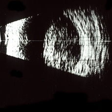 Choroidal melanoma; B-scan
May 2 2013 by Henry J. Kaplan, MD
B-scan of a choroidal melanoma shows dome shaped lesion #1.
Condition/keywords: B scan ultrasound
-
 Radiation retinopathy
May 2 2013 by Henry J. Kaplan, MD
Radiation maculopathy after brachytherapy of a choroidal melanoma; notice the telangiectatic changes and retinal exudation.
Condition/keywords: radiation maculopathy, radiation retinopathy
-
 Choroidal melanoma case 2 image 2
Jan 11 2013 by Alex P. Hunyor, MD
Large right choroidal melanoma - color image 2 of 2
Condition/keywords: melanoma
-
 Amelanotic choroidal melanoma
Dec 19 2012 by Eric A. Postel, MD
-
 Diffuse Choroidal Melanoma OD
Aug 24 2012 by John S. King, MD
AF
Photographer: Kristin Konecki, OcuSight Eye Care Center, Rochester, NY
-
 Choroidal Melanoma
Jan 30 2019 by Karen Panzegrau
Ultra-wide field optos image of a 27-year-old male patient who presented with loss of vision for about 6-8 weeks. Previous choroidal nevus seen. Recommended annual monitoring. No exam for since 10/2014. Brachytherapy vs enucleation was discussed. Brachytherapy was decided as treatment. Full metastatic work up is being performed.
Photographer: Karen Panzegrau
Imaging device: Optos
Condition/keywords: choroidal nevus, exudative retinal detachment, malignant neoplasm of eye, Optos, ultra-wide field imaging
-
 Amelanotic Choroidal Melanoma
Apr 12 2019 by David L Kilpatrick, MD
Fundus photograph of a 69-year-old male with an amelanotic choroidal melanoma and corresponding exudative retinal detachment. Transvitreal biopsy was performed at the time of radioactive I-125 plaque placement. The genetic expression profile revealed a Class 1A, PRAME negative tumor.
Photographer: Retina Consultants of Alabama, P. C.
Imaging device: Optos
Condition/keywords: amelanotic melanoma
-
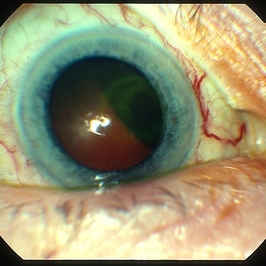
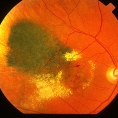
 Orange Pigment Overlying a Lesion Suspicious for a Choroidal Melanoma
Jan 16 2019 by John S. King, MD
76-year-old white male saw his eye doctor with a three week complaint of photopsias and a shadow in his vision. Found to have a 10.5/12.5/2.5 (medium reflectivity) pigmented, choroidal mass associated with SRF and orange pigment (hyper-autofluorescence of lipofuscin), and without drusen or halo. See photo
Photographer: Stacey Coleman
Imaging device: Topcon 50
Condition/keywords: lipofuscin, orange pigment
-
 Choroidal Melanoma With Radiation Retinopathy
Jul 8 2013 by Jason S. Calhoun
Patient came with follow up on choroidal melanoma. Right eye that was treated back in June of 2009 with a radioactive implant. Vein occlusion is also present with VA - hand motion. Hemorrhages visible with hard exudates from the radiation retinopathy.
Photographer: Jason S. Calhoun, Department of Ophthalmology, Mayo Clinic Jacksonville, Florida
Condition/keywords: radiation retinopathy
-
 Malignant Choroidal Melanoma
Dec 4 2015 by Kathy Karsten, COT
Malignant choroidal melanoma and branch retinal vein occlusion in 69-year-old male.
Photographer: Kathy Karsten, COT
Imaging device: Topcon TRC-50 DX
 Loading…