Search results (86 results)
-
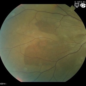
---thumb.JPG/image-square;max$300,300.ImageHandler) Choroidal Hemorrhage
Choroidal Hemorrhage
Jul 11 2013 by Jason S. Calhoun
Choroidal hemorrhage.
Photographer: Jason S. Calhoun, Department of Ophthalmology, Mayo Clinic Jacksonville, Florida
Condition/keywords: choroidal hemorrhage
-
 Choriodal Rupture - AW 001 - Initial Presentation
Mar 11 2013 by Suber S. Huang, MD, MBA, FASRS
40-year-old male sustained blunt trauma OD with orbital fracture and choroidal rupture subjacent to inferior arcade with blood, subretinal fluid, and exudate extending to fovea with CF 2 feet at presentation 9-10-12. On followup 3-11-12, vision improved to 20/400 with resolution of hemorrhage, normal OCT, but speckling of foveal RPE and PMB consistent with damage.
Photographer: Mark Harrod
Condition/keywords: autofluorescence imaging, blunt trauma, choroidal hemorrhage, choroidal rupture, orbital fracture, retinal hemorrhage, submacular hemorrhage
-
 Choriodal Rupture - AW 002 - 6 Month F/U
Mar 11 2013 by Suber S. Huang, MD, MBA, FASRS
40-year-old male sustained blunt trauma OD with orbital fracture and choroidal rupture subjacent to inferior arcade with blood, subretinal fluid, and exudate extending to fovea with CF 2 feet at presentation 9-10-12. On follow up 3-11-12, vision improved to 20/400 with resolutionof hemorrhage, normal OCT, but speckling of foveal RPE and PMB consistent with damage.
Photographer: Mark Harrod
Condition/keywords: autofluorescence imaging, blunt trauma, choroidal hemorrhage, choroidal rupture, orbital fracture, retinal hemorrhage, submacular hemorrhage
-
 Choriodal Rupture 004 - Fundus Autoflurescence - 6 Month Follow Up
Mar 11 2013 by Suber S. Huang, MD, MBA, FASRS
40-year-old male sustained blunt trauma OD with orbital fracture and choroidal rupture subjacent to inferior arcade with blood, subretinal fluid, and exudate extending to fovea with CF 2 feet at presentation 9-10-12. On follow up 3-11-12, vision improved to 20/400 with resolutionof hemorrhage, normal OCT, but speckling of foveal RPE and PMB consistent with damage.
Photographer: Mark Harrod
Condition/keywords: autofluorescence imaging, blunt trauma, choroidal hemorrhage, choroidal rupture, orbital fracture, retinal hemorrhage, submacular hemorrhage
-
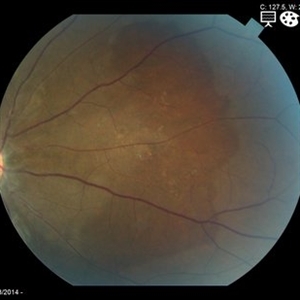
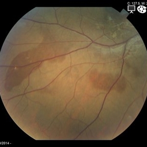 Choroidal Hemorrhage from Hypertensive Choroidopathy
May 19 2014 by Charline Boente
Fundus photograph of a 21-year-old African-American female with HTN and end-stage renal disease from IgA nephropathy.
Photographer: Mark Harrod, University Hospitals Eye Institute, Case Western Reserve University
Condition/keywords: hypertensive choroidopathy
-
 Choroidal Hemorrhage, Subretinal Hemorrhage
Dec 18 2017 by Nichole Lewis
Choroidal hemorrhage, Subretinal Hemorrhage, wet macular degeneration,
Photographer: Nichole Lewis
Condition/keywords: choroidal hemorrhage, choroidal neovascularization (CNV), exudative age-related macular degeneration, subretinal hemorrhage, wet age-related macular degeneration (wet AMD)
-
 Choroidal Hemorrhage
Mar 29 2013 by Henry J. Kaplan, MD
Traumatic choroidal hemorrhage with possible underlying choroidal ruptures.
Condition/keywords: choroidal hemorrhage
-
 Choriodal Rupture - 003 - 6 Month Follow Up
Mar 11 2013 by Suber S. Huang, MD, MBA, FASRS
40 -year-old male sustained blunt trauma OD with orbital fracture and choroidal rupture subjacent to inferior arcade with blood, subretinal fluid, and exudate extending to fovea with CF 2 feet at presentation 9-10-12. On followup 3-11-12, vision improved to 20/400 with resolutionof hemorrhage, normal OCT, but speckling of foveal RPE and PMB consistent with damage.
Photographer: Mark Harrod
Condition/keywords: autofluorescence imaging, blunt trauma, choroidal hemorrhage, choroidal rupture, orbital fracture, retinal hemorrhage, submacular hemorrhage
-
 Scleritis
Jul 16 2014 by John S. King, MD
Keratoscleritis with suprachoroidal hemorrhage in an elderly male with history of cataract surgery.
Photographer: URMC
Condition/keywords: keratitis, scleritis, suprachoroidal hemorrhage
-
 Choroidal Hemorrhage from Hypertensive Choroidopathy
May 19 2014 by Charline Boente
Fundus photograph of a 21-year-old African-American female with HTN and end-stage renal disease from IgA nephropathy.
Photographer: Mark Harrod, University Hospitals Eye Institute, Case Western Reserve University
Condition/keywords: hypertensive choroidopathy
-
 Vitreous & Subretinal Hemorrhage in Polypoidal Choroidal Vasculopathy
Dec 10 2012 by Yale L. Fisher, MD
Type 1 CNV (polypoidal choroidal vasculopathy) with vitreous hemorrhage and suprachoroidal hemorrhage.
Condition/keywords: polypoidal choroidal vasculopathy (PCV), suprachoroidal hemorrhage, video, vitreous hemorrhage
-
 Massive Suprachoroidal & Subretinal Hemorrhage in IPCV
Oct 7 2014 by Mallika Goyal, MD
53-year-old lady with bilateral PCV had limited nasal RPE detachment in left eye . No response to anti-VEGF therapy. Treated with PDT. This is 3 days post-PDT exudative retinal detachment.
Photographer: Mallika Goyal, MD, Apollo Health City, Jubilee Hills, Hyderabad, India
Condition/keywords: suprachoroidal hemorrhage
-
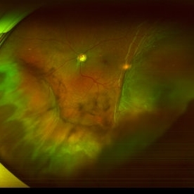 Suprachoroidal Hemorrhage
Sep 2 2020 by Rinal Pandit
Fundus photograph of left eye of a 56-year-old female with primary angle closure glaucoma showing massive hemorrhagic choroidal detachment that developed following trabeculectomy surgery. Suprachoroidal hemorrhage is defined as the accumulation of blood within the potential space between the choroid and sclera, with the source of the blood being the long or short posterior ciliary artery. Delayed suprachoroidal hemorrhage (DSHC) remains one of the most dreaded and sight threatening complications of glaucoma filtration surgery. The risk factors include old age, hypertension, high myopia, arteriosclerosis, chronically elevated IOP, sudden hypotony, trauma, aphakia/pseudophakia, prior vitrectomy, history of 5 FU injections and anti-platelet agents. The incidence of postoperative SCH after trabeculectomy varies between 0.6%- 1.4%. DSCH after surgery varies considerably in severity but is generally characterized by the sudden onset of severe pain, decreased vision, and a shallow anterior chamber usually associated with raised intraocular pressure. B-scan ultrasonography can help to distinguish serous from hemorrhagic choroidals.Suprachoroidal hemorrhages appear as dome-shaped elevations of the retina with increased echo densities that are often heterogeneous and within the suprachoroidal space. Choroidal effusions appear as dome-shaped elevations with hypoechoic suprachoroidal space. The first step in the management is the timely diagnosis. Medical management includes oral and topical antiglaucoma drugs to lower IOP, oral and topical steroids to control inflammation and topical cycloplegics and oral analgesics to tackle pain. Serial ultrasound B scans of the affected eye should be performed in order to monitor progression of the SCH and help determine apposition, height, and liquefaction of the SCH. Indications of surgical drainage include non resolution with medical management,concurrent retinal detachment, central retinal apposition (kissing choroidals) and incarceration of vitreous in the wound site. The ideal time of drainage is between 7-14 days depending upon clot lysis. The prognosis of both intraoperative and postoperative SCH is poor. An overwhelming majority of patients do not achieve pre-hemorrhage visual acuity and most do not recover to a visual acuity of 20/200 or better. The major determinants of good or bad visual outcomes of SCH’s are preoperative visual acuity and retinal detachment at the time of hemorrhage, respectively.
Imaging device: OPTOS,Ultra wide field retinal imaging system
Condition/keywords: suprachoroidal hemorrhage, trabeculectomy, ultra-wide field imaging
-
 Choroidal Hemorrhage from Hypertensive Choroidopathy
May 19 2014 by Charline Boente
Fundus photograph of a 21-year-old African-American female with HTN and end-stage renal disease from IgA nephropathy.
Photographer: Mark Harrod, University Hospitals Eye Institute, Case Western Reserve University
Condition/keywords: hypertensive choroidopathy
-
 Scleritis
Jul 16 2014 by John S. King, MD
Keratoscleritis with suprachoroidal hemorrhage in an elderly male with history of cataract surgery.
Photographer: URMC
Condition/keywords: keratitis, scleritis, suprachoroidal hemorrhage
-
 Scleritis
Jul 16 2014 by John S. King, MD
Keratoscleritis with suprachoroidal hemorrhage in an elderly male with history of cataract surgery.
Photographer: URMC
Condition/keywords: keratitis, scleritis, suprachoroidal hemorrhage
-
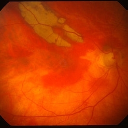
 Choroidal And Subretinal Hemorrhage
Jul 11 2013 by Jerald A. Bovino, MD
No history.
Condition/keywords: choroidal hemorrhage, subretinal hemorrhage
-
 Choroidal Hemorrhage from Hypertensive Choroidopathy
May 19 2014 by Charline Boente
Fundus photograph of a 21-year-old African-American female with HTN and end-stage renal disease from IgA nephropathy.
Photographer: Mark Harrod, University Hospitals Eye Institute, Case Western Reserve University
Condition/keywords: hypertensive choroidopathy
-
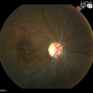
 Superchoroid Hemorrhage
Feb 12 2015 by H. Michael Lambert, MD
A-scan shows choroidal detachment with hemorrhage.
Condition/keywords: choroidal detachment, choroidal hemorrhage
-
 Massive Suprachoroidal & Subretinal Hemorrhage in IPCV
Oct 7 2014 by Mallika Goyal, MD
53-year-old lady with bilateral PCV had extensive lesions right eye, limited nasal RPE detachment in left eye. No response to anti-VEGF therapy with gradual worsening status. Treated with PDT and Avastin. This is pre-PDT right eye fundus photograph.
Photographer: Mallika Goyal, MD, Apollo Health City, Jubilee Hills, Hyderabad, India
Condition/keywords: suprachoroidal hemorrhage
-
 Superchoroid Hemorrhage
Feb 12 2015 by H. Michael Lambert, MD
Choroidal detachment.
Condition/keywords: choroidal detachment, choroidal hemorrhage
-
 Scleritis
Jul 16 2014 by John S. King, MD
Keratoscleritis with suprachoroidal hemorrhage in an elderly male with history of cataract surgery.
Photographer: URMC
Condition/keywords: keratitis, scleritis, suprachoroidal hemorrhage
-
 Massive Suprachoroidal & Subretinal Hemorrhage in IPCV
Oct 7 2014 by Mallika Goyal, MD
53-year-old lady with bilateral PCV had limited nasal RPE detachment in left eye. No response to anti-VEGF therapy. Treated with PDT. This is 3 days post-PDT exudative retinal detachment.
Photographer: Mallika Goyal, MD, Apollo Health City, Jubilee Hills, Hyderabad, India
Condition/keywords: suprachoroidal hemorrhage
-
 Scleritis
Jul 16 2014 by John S. King, MD
Keratoscleritis with suprachoroidal hemorrhage in an elderly male with history of cataract surgery.
Photographer: URMC
Condition/keywords: keratitis, scleritis, suprachoroidal hemorrhage
-
 Massive Suprachoroidal & Subretinal Hemorrhage in IPCV
Oct 7 2014 by Mallika Goyal, MD
53-year-old lady with bilateral PCV had limited nasal RPE detachment in left eye . No response to anti-VEGF therapy. Treated with PDT. This is 3 days post-PDT exudative retinal detachment.
Photographer: Mallika Goyal, MD, Apollo Health City, Jubilee Hills, Hyderabad, India
Condition/keywords: suprachoroidal hemorrhage
 Loading…