Search results (12 results)
-
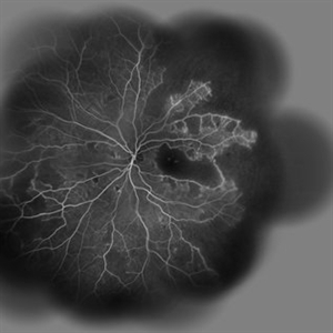
 Macular Ischemia
Aug 23 2012 by Gerardo Garcia-Aguirre, MD
Fluorescein angiogram of a right eye with proliferative diabetic retinopathy that underwent panretinal photocoagulation showing vast areas of capillary closure that involve the macular area.
Photographer: Noemí Hernández, Asociación para Evitar la Ceguera en México
Condition/keywords: macular ischemia
-
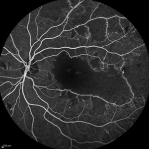
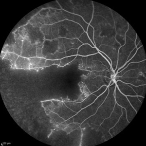 Idiopathic Occlusive Retinal Vasculitis (Late Stage)
May 31 2014 by Hamid Ahmadieh, MD
Late phase FA image of the right eye of a 28-year-old woman with idiopathic occlusive retinal vasculitis 6 months after the onset.
Photographer: Solmaz Shahmohammad, Negah Eye Center, Tehran
Imaging device: Heidelberg Spectralis
Condition/keywords: capillary closure, fluorescein leakage, macular infarction
-
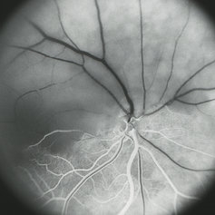
 Amikacin Retinal Toxicity
Sep 26 2012 by Jose Dalma-Weiszhausz, MD
Extensive capillary closure after intravitreal amikacin injection.
Photographer: José Dalma, MD, Dalma & Asoc. Mexico City, Mexico
Condition/keywords: Amikacin retinal toxicity
-
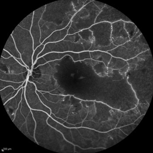
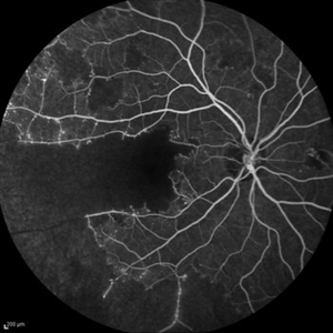 Idiopathic Occlusive Retinal Vasculitis (Late Stage)
May 31 2014 by Hamid Ahmadieh, MD
Late phase FA image of the right eye of a 28-year-old woman with idiopathic occlusive retinal vasculitis 6 months after the onset.
Photographer: Solmaz Shahmohammad, Negah Eye Center, Tehran
Imaging device: Heidelberg Spectralis
Condition/keywords: capillary closure, macular infarction
-
 Fluoroangiogram of Amikacin Retinal Toxicity, Macular Capillary Closure
Sep 26 2012 by Jose Dalma-Weiszhausz, MD
Extensive capillary closure after intravitreal amikacin injection.
Photographer: José Dalma MD, Dalma & Asoc. Mexico City, Mexico
Condition/keywords: Amikacin retinal toxicity, capillary closure
-
 Idiopathic Occlusive Retinal Vasculitis (Late Stage)
May 31 2014 by Hamid Ahmadieh, MD
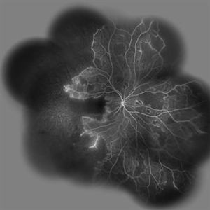
Wide-field FA image of the left eye of a 28-year-old woman with idiopathic occlusive retinal vasculitis 6 months after the onset.
Photographer: Elham Salehi, Negah Eye Center, Tehran
Imaging device: Heidelberg Spectralis
Condition/keywords: capillary closure, macular infarction
-
 Idiopathic Occlusive Retinal Vasculitis (Late Stage)
May 31 2014 by Hamid Ahmadieh, MD
Wide- field image of the right eye of a 28-year-old woman with idiopathic occlusive retinal vasculitis 6 months after the onset.
Photographer: Solmaz Shahmohammad, Negah Eye Center, Tehran
Imaging device: Heidelberg Spectralis
Condition/keywords: capillary closure, macular infarction
-
 Idiopathic Occlusive Retinal Vasculitis (Late Stage)
May 31 2014 by Hamid Ahmadieh, MD
Mid- venous phase FA image of the left eye of a 28-year-old woman with idiopathic occlusive retinal vasculitis leading to extensive capillary closure macular infarction and collaterals 6 month after the onset.
Photographer: Elham Salehi, Negah Eye Center, Tehran
Imaging device: Heidelberg Spectralis
Condition/keywords: capillary closure
-
 Idiopathic Occlusive Retinal Vasculitis (Late Stage)
May 31 2014 by Hamid Ahmadieh, MD
Late phase FA image of the left eye of a 28-year-old woman with idiopathic occlusive retinal vasculitis 6 months after the onset.
Photographer: Elham Salehi, Negah Eye Center, Tehran
Imaging device: Heidelberg Spectralis
Condition/keywords: capillary closure
-
 Normal
Jul 22 2013 by Howard Schatz, MD
Leak, PE, BM, CC.
Condition/keywords: BM, capillary closure, leakage, normal eye, pigment epithelium
-
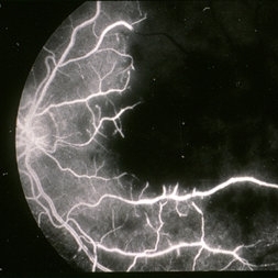
 Hemi-CRAO
Mar 26 2019 by Gary R. Cook, MD, FACS
Mid-phase (laminar venous return) fluorescein angiogram image of an embolic superior hemi-CRAO showing marked delay in filling of the superior retinal arteriolar and venous vasculature and total loss of the retinal capillary bed in the superior hemisphere OD.
Condition/keywords: capillary closure, capillary nonperfusion, central retinal artery occlusion (CRAO), FA mid phase, fluorescein angiogram (FA)
-
 Idiopathic retinal vasculitis, aneurysms and neuroretinitis
Apr 24 2022 by Aniruddha K Agarwal, MD
Ultra-wide field fundus fluorescein angiography (FFA) of the left eye from an asymptomatic, healthy 33-year-old woman who was referred to the retina clinic from a refractive surgery unit due to the presence of vascular anomalies and hard exudates in both eyes. FFA revealed the characteristic sacular aneurysms at the bifurcation of retinal arterioles in the posterior pole, together with microvascular anomalies and capillary closure peripherally.
Photographer: Julio J GONZALEZ-LOPEZ, MD, PhD, FEBO and Teresa GONZALEZ-LOMAS, RN
Imaging device: Optos California
Condition/keywords: IRVAN Syndrome, IUSG, neuroretinitis, retinal vasculitis, uveitis
 Loading…