Search results (23 results)
-
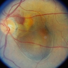
---thumb.JPG/image-square;max$300,300.ImageHandler) Retinal Pigment Epithelial Detachment With No Subretinal Fluid
Retinal Pigment Epithelial Detachment With No Subretinal Fluid
Jun 29 2013 by Jason S. Calhoun
A 38-year-old male who comes in with blurred vision in the left eye. VA is 20/30. Noticed a defect inferior of his central vision. Did an fluorescein angiogram to determine an RPE with no sub retinal fluid. Also OCT confirms. Patient was injected with Avastin.
Photographer: Jason S. Calhoun, Mayo Clinic Jacksonville, Florida
Imaging device: TOPCON TRC 50-EX
Condition/keywords: central serous retinopathy (CSR), retinal pigment epithelium (RPE) detachment
-
 Optic Neuritis With Suspected Harada's Disease
Jun 27 2013 by Jason S. Calhoun
Young female patient came in with red and irritated eyes which were sensitive to the touch. Patient also had vision loss in both eyes. VA was 20/80, right eye, 20/60, left eye. Fundiscopic exam shows sub retinal fluid in the right eye surrounding the optic nerve. Patient will start steroid therapy to reduce any other inflammation that is active
Photographer: Jason S. Calhoun, Mayo Clinic Jacksonville, Florida
Imaging device: TOPCON TRC 50-EX
Condition/keywords: Harada's disease, optic neuritis
-
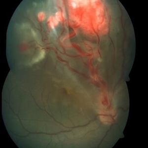 Retinal Capillary Hemangioblastoma
Oct 6 2015 by Pukhraj P Rishi, MBBS, MS, DO, FRCS, FRCSEd, FASRS, FACS
Fundus photograph of an 18-year-old Asian Indian male with multiple retinal capillary hemangiomas with sub retinal fluid.
Photographer: M S KRISHNA
Imaging device: Zeiss FF4
Condition/keywords: retinal angioma, retinal capillary hemangioma, tumor, Von Hippel-Lindau
-
 Horseshoe Tear
Sep 17 2015 by Jason S. Calhoun
Horseshoe tear with sub retinal fluid present superior temporal in the left eye.
Photographer: Jason Calhoun, Mayo Clinic, Department of Ophthalmology
Imaging device: OPTOS 200TX
-
 Melanoma with Subretinal Fluid
Jul 14 2013 by Jason S. Calhoun
52-year-old female who comes in for eye exam. Fundus exam shows yellowish elevated spot superior at 12-o'clock in the right eye. FA shows sub retinal fluid with melanoma in the right eye.
Photographer: Jason S. Calhoun, Department of Ophthalmology, Mayo Clinic Jacksonville, Florida
Imaging device: TOPCON TRC 50-EX
Condition/keywords: melanoma, subretinal fluid
-
---thumb.JPG/image-square;max$300,300.ImageHandler) Retinal Pigment Epithelial Detachment With No Subretinal Fluid
Retinal Pigment Epithelial Detachment With No Subretinal Fluid
Jun 29 2013 by Jason S. Calhoun
A 38-year-old male who comes in with blurred vision in the left eye. VA is 20/30. Notices a defect inferior of his central vision. Did an fluorescein angiogram to determine an RPE with no sub retinal fluid. Also OCT confirms. Patient was injected with Avastin.
Photographer: Jason S. Calhoun, Mayo Clinic Jacksonville, Florida
Imaging device: TOPCON TRC 50-EX
Condition/keywords: central serous retinopathy (CSR), retinal pigment epithelium (RPE) detachment
-
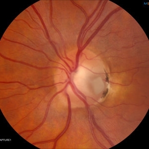 Optic Nerve Pit With Sub-Retinal Fluid
Sep 17 2015 by Jason S. Calhoun
Young female with blurred vision in the left eye. Fundus photograph shows optic nerve pit adjacent to the macula where there is sub retinal fluid visible.
Photographer: Jason Calhoun, Mayo Clinic, Department of Ophthalmology
Imaging device: TOPCON-TRC50EX
Condition/keywords: congenital optic nerve pit
-
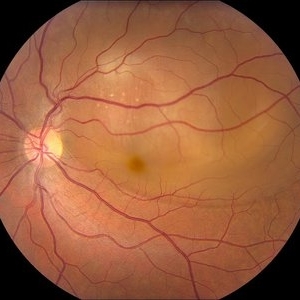 Severe CSR
Jul 14 2013 by Jason S. Calhoun
A 38-year-old male came in with blurred vision in the left eye. VA is 20/30. A defect inferior of his central vision was noticed and a fluorescein angiogram was done to determine an RPE with no sub retinal fluid. Also OCT confirms. Patient was injected with Avastin.
Photographer: Jason S. Calhoun, Department of Ophthalmology, Mayo Clinic Jacksonville, Florida
Imaging device: TOPCON TRC 50-EX
Condition/keywords: central serous retinopathy (CSR)
-
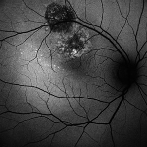
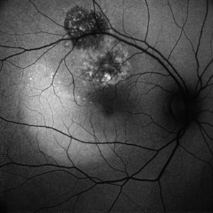
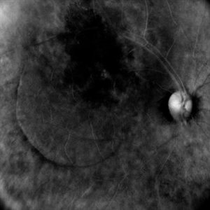
---thumb.JPG/image-square;max$300,300.ImageHandler) Montage of CSR (FAF)
Montage of CSR (FAF)
Jul 14 2013 by Jason S. Calhoun
A 38-year-old male came in with blurred vision in the left eye. VA is 20/30. A defect inferior of his central vision was noticed and a fluorescein angiogram was done to determine an RPE with no sub retinal fluid. Also OCT confirms. Patient was injected with Avastin.
Photographer: Jason S. Calhoun, Department of Ophthalmology, Mayo Clinic Jacksonville, Florida
Imaging device: TOPCON TRC 50-EX
Condition/keywords: central serous retinopathy (CSR)
-
 Optic Nerve Pit with Sub-Retinal Fluid
Sep 17 2015 by Jason S. Calhoun
Young female with blurred vision in the left eye. Fundus photograph shows optic nerve pit adjacent to the macula where there is sub retinal fluid visible.
Photographer: Jason Calhoun, Mayo Clinic, Department of Ophthalmology
Imaging device: TOPCON-TRC50EX
Condition/keywords: congenital optic nerve pit
-
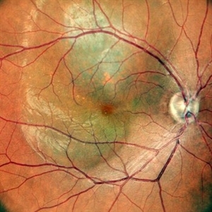
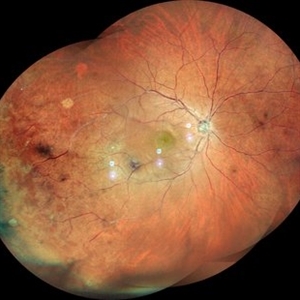
---thumb.JPG/image-square;max$300,300.ImageHandler) Montage of CSR (Color)
Montage of CSR (Color)
Jul 14 2013 by Jason S. Calhoun
A 38-year-old male who came in with blurred vision in the left eye. VA is 20/30. A defect inferior of his central vision was noticed and a fluorescein angiogram was done to determine an RPE with no sub retinal fluid. Also OCT confirms. Patient was injected with Avastin.
Photographer: Jason S. Calhoun, Department of Ophthalmology, Mayo Clinic Jacksonville, Florida
Imaging device: TOPCON TRC 50-EX
Condition/keywords: central serous retinopathy (CSR)
-
 Chronic Retinal Detachment after Pneumatic Retinopexy
Jan 8 2022 by Parnian Arjmand, MD, MSc, FRCSC, DABO
This is a fundus photo in the eye of a young phakic patent who presented with a 6 month history of "difficulty seeing at night" and subjective nasal "blurriness" in the left eye. There was a chronic temporal RD, OS, extending to the arcades (Mac on). This photo is week 1 s/p Pneumatic retinopexy with SF6 gas and laser retinopexy to temporal breaks (6 holes, lattice); no PVD. As you can see, there is a "bleb" of viscous schlieren given the chronic nature of this RD that persist posterior to the breaks and temporal to the macula. This type of sub retinal fluid may take months to years to resorb.
Condition/keywords: chronic retinal detachment, pneumatic retinopexy
-
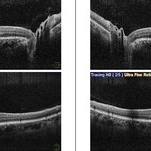
 Persistence of Sub Retinal Fluid Post Retinal Reattachment Surgery
Sep 14 2021 by Ogugua Ndubuisi Okonkwo, MD, FRCS (Edin), FASRS
Postoperative Optical Coherence Tomography (OCT) of the right eye of a 35-year-old male showing persistence of subretinal fluid in the original area of the retinal detachment.
Photographer: Oreoluwa , Eye Foundation Hospital, Lagos
Imaging device: Optovue Avanti RTVue
Condition/keywords: re-attached retinal detachment (RRD), subretinal fluid
-
 Peripapillary Choroidal Neovascular Membrane (CNVM)
Sep 26 2023 by Ben Serar
Fundus photograph showing Peripapillary Choroidal Neovascular Membrane (CNVM) with subretinal bleed and surrounding sub retinal fluid.
Condition/keywords: Peripapillary Choroidal Neovascular Membrane (CNVM)
-
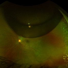
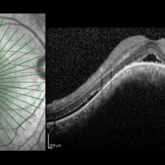
Peripheral Retinal Hole with OCT Co-localization
Sep 26 2023 by Bradley T. Smith, MD, FASRS
Peripheral asymptomatic atrophic retinal hole with OCT co localization demonstrating small cuff of sub retinal fluid. Near infrared imaging shows hyper reflectivity through hole.
Condition/keywords: atrophic hole, lattice degeneration, OCT
-
 CHOROIDAL HEMANGIOMA
Nov 21 2022 by Akansha Sharma
GREEN-AUTOFLUORESCENCE IMAGE OF A 32 YEAR OLD MALE WITH CHOROIDAL HEMANGIOMA WITH SUB RETINAL FLUID
Photographer: Dr. Akansha Sharma-Retina Foundation, Ahmedabad
Condition/keywords: choroidal hemangioma, subretinal fluid
-
 CHOROIDAL HEMANGIOMA
Nov 21 2022 by Akansha Sharma
COLOU FUNDUS IMAGE OF A 32 YEAR OLD MALE WITH CHOROIDAL HEMANGIOMA WITH SUB RETINAL FLUID
Photographer: Dr. Akansha Sharma-Retina Foundation, Ahmedabad
Condition/keywords: choroidal hemangioma, subretinal fluid
-
 CHOROIDAL HEMANGIOMA
Nov 21 2022 by Akansha Sharma
BLUE-AUTOFLUORESCENCE IMAGE OF A 32 YEAR OLD MALE WITH CHOROIDAL HEMANGIOMA WITH SUB RETINAL FLUID
Photographer: Dr. Akansha Sharma-Retina Foundation, Ahmedabad
Condition/keywords: choroidal hemangioma, subretinal fluid
-
 CHOROIDAL HEMANGIOMA
Nov 21 2022 by Akansha Sharma
RETRO IMAGE OF A 32 YEAR OLD MALE WITH CHOROIDAL HEMANGIOMA WITH SUB RETINAL FLUID
Photographer: Dr. Akansha Sharma-Retina Foundation, Ahmedabad
Condition/keywords: choroidal hemangioma, subretinal fluid
-
 Choroidal Mass
Mar 4 2024 by ANKIT JAIN
RE color photo montage of right eye of 48 year old with sub retinal hemorrhage with sub retinal fluid at level of fovea.
Photographer: Dr Ankit Jain
Imaging device: MIRANTE
Condition/keywords: macroaneurysm, retinal arterial macroaneurysm
-
 OCT in Adult Vitelliform Dystrophy
Jun 25 2024 by Tejaswita Verma
OCT image of a 62 year old female with 6/12 vision in both eyes showing sub retinal fluid with RPE granularity s/o Adult vitelliform macular dystrophy.
Photographer: DR. TEJASWITA VERMA
Imaging device: MIRANTE
Condition/keywords: adult vitelliform dystrophy, optical coherence tomography (OCT)
-
OCT Video Imaging of Left Eye Age Related Macular Degeneration
Jan 6 2025 by Kavitha Duraipandi, MD DNB FICO FRCS
Left eye OCT macula shows various biomarkers like PED, sub retinal fluid, sub retinal hyper reflective material and hyper reflective foci suggestive of wet age-related macular degeneration.
Condition/keywords: OCT biomarkers, wet age-related macular degeneration (wet AMD)
-
 Choroidal Hemangioma
Jun 18 2025 by Moazzam Parvez
An OCT image of a 42 year old man presenting with a vision of 20/80 and complaining of distortion. OCT reveals serous retinal detachment with RPE alteration and disruption of outer retinal layers.
Photographer: Moazzam Parvez , Netralayam , Kolkata
Imaging device: Heidelberg Spectralis
Condition/keywords: Choroidal Hemangioma, Sub retinal fluid, tumor
 Loading…