Search results (13 results)
-
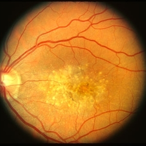
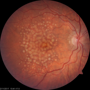
 Reticular Drusen, Doyne's Honeycomb Retinal Dystrophy, Malattia Leventinese, Familial Dominant Drusen
Feb 22 2018 by Nichole Lewis
Reticular Drusen, Doyne's Honeycomb Retinal Dystrophy, Malattia Leventinese, Familial Dominant Drusen
Photographer: Nichole Lewis
Condition/keywords: Doyne's Honeycomb, Familial Dominant Drusen, Malattia Leventinese, reticular drusen
-
 Malattia Leventinese 2 - color LE
Jan 11 2013 by Alex P. Hunyor, MD
Malattia levantinese - left eye. Asymptomatic patient with 20/25 vision OU.
Condition/keywords: Malattia Leventinese
-
 Reticular Drusen, Doyne's Honeycomb Retinal Dystrophy, Malattia Leventinese, Familial Dominant Drusen
Feb 22 2018 by Nichole Lewis
Reticular Drusen, Doyne's Honeycomb Retinal Dystrophy, Malattia Leventinese, Familial Dominant Drusen
Photographer: Nichole Lewis
Condition/keywords: Doyne's Honeycomb, Familial Dominant Drusen, Malattia Leventinese, reticular drusen
-
 Malattia Leventinese 1 - colour RE
Jan 11 2013 by Alex P. Hunyor, MD
Malattia Leventinese - right eye.
Condition/keywords: Malattia Leventinese
-
 Malattia Leventinese 2 - color RE
Jan 11 2013 by Alex P. Hunyor, MD
Malattia leventinese - right eye. Asymptomatic patient with 20/25 vision OU.
Condition/keywords: Malattia Leventinese
-
 Malattia Leventinese 1 - colour LE
Jan 11 2013 by Alex P. Hunyor, MD
Malattia leventinese - left eye - complicated by CNV (note subretinal haemorrhage).
Condition/keywords: Malattia Leventinese
-
 Colloidal Drusen
Nov 6 2017 by Ian C Reddie, LLB (QUT), MBBS (Qld), FRANZCO, FASRS
Fundus photograph of 45-year-old woman showing large colloidal drusen. Previously misdiagnosed as malattia leventinese.
Photographer: Dr Ian C Reddie, North Queensland Retina, Townsville, Queensland, Australia
Condition/keywords: colloidal drusen, drusen
-
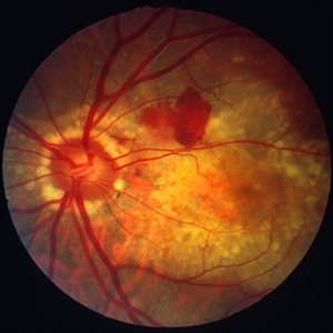
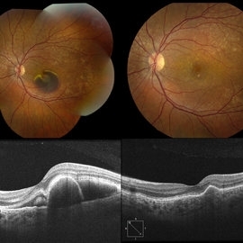 Submacular Hemorrhage Due to Malattia Leventinese
Dec 29 2018 by Darin R. Goldman, MD
Fundus photograph and OCT of a 37-year-old female with a presumed diagnosis of Malattia Leventinese. One of the more characteristic features of this condition, as seen with this case, is the radiating pattern of drusen and particularly their location nasal to the optic nerve. There is a submacular hemorrhage due to a secondary CNV, which resolved after a series of anti-VEGF therapy with ranibizumab. The followup images (right side) are from 3.5 months after presentation where her VA recovered to 20/20.
Photographer: Robin Lapointe, CRA, Retina Group of Florida
Imaging device: Topcon TRC 50 DX
Condition/keywords: Doyne's Honeycomb, familial drusen, Malattia Leventinese, submacular hemorrhage
-
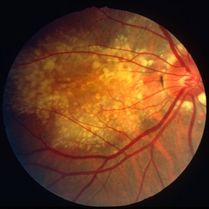
 Doyne Honeycomb Retinal Dystrophy
Sep 29 2020 by Navneet Mehrotra, DNB
Left eye fundus photograph of a 36-year-old female with decreased vision both eyes for six months. Father also had a similar retinal disorder.
Photographer: Dr Navneet Mehrotra
Imaging device: TRC- NW8F
Condition/keywords: Doyne's Honeycomb, drusen, Malattia Leventinese
-
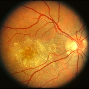
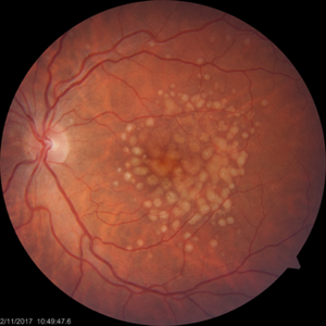
 Malattia Leventinese
Jul 19 2019 by Anfisa Ayalon, MD
36-years-old male with both eyes Familial Dominant Drusen. Snellen chart visual acuity in both eyes-20/20.
Photographer: Anfisa Ayalon, MD., Meir Medical Center, Kfar Saba, Israel.
Condition/keywords: drusen, drusenoid deposit, familial drusen, Malattia Leventinese
-
 Colloidal Drusen
Nov 6 2017 by Ian C Reddie, LLB (QUT), MBBS (Qld), FRANZCO, FASRS
Fundus photograph of 45-year-old woman showing large colloidal drusen. Previously misdiagnosed as malattia leventinese.
Photographer: Dr Ian C Reddie, North Queensland Retina, Townsville, Queensland , Australia
Condition/keywords: colloidal drusen, drusen
-
 Doyne Honeycomb Retinal Dystrophy
Sep 29 2020 by Navneet Mehrotra, DNB
Right eye fundus photograph of a 36-year-old female with decreased vision both eyes for six months. Father also had a similar retinal disorder.
Photographer: Dr Navneet Mehrotra, Retina Care, Ahmedabad
Imaging device: TRC- NW8F
Condition/keywords: Doyne's Honeycomb, familial drusen, Malattia Leventinese
-
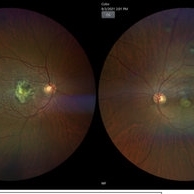 A rare case of a 45-year-old male with choroidal neovascular membrane in Familial Dominant Drusen (Doyne Honeycomb Drusen) in both eyes treated with intravitreal injections.
Nov 30 2022 by SHRADDHA ASHOK CHANDORKAR, DNB DO
A 45-year-old man presented with diminution of vision in both eyes with metamorphopsia, which was painless and gradually progressive in nature. BCVA at presentation were 6/40 and 6/36 for the right and left eye respectively. Anterior segment examination of both eyes was unremarkable. IOP were within normal limits. Fundus examination showed bilateral numerous yellowish white round closely spaced lesions extending radially from the vascular arcades till the periphery associated with an elevated grayish macular choroidal neovascular membrane (CNV) with multiple drusen in the macular area and posterior pole. Impression was Familial Dominant Drusen (Doyne Honeycomb Drusen) associated with CNVM, both eyes. Color fundus photograph and autofluorescence showed Familial Dominant Drusen with CNVM. Subsequently , the patient underwent periodic intravitreal injections of Ranibizumab in both eyes under guarded visual prognosis, for which he tolerated well.
Photographer: NATIONAL INSTITUTE OF OPHTHALMOLOGY, PUNE
Imaging device: ZEISS CLARUS
Condition/keywords: choroidal neovascular membrane (CNVM), Doyne's Honeycomb, FAMILIAL DOMINANT DRUSEN, IMIM (Online Mendelian Inheritance in Man), intravitreal injection, Malattia Leventinese
 Loading…