Search results (11 results)
-
 Gardner Syndrome
Dec 12 2018 by John S. King, MD
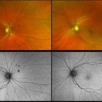
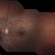
66-year-old white male with Gardner Syndrome (colon resection in 1991), who has two children with Gardner Syndrome, presented to Dr. Zocchi with an RD in the fellow eye that was successfully repaired with a pneumatic retinopexy. Currently 20/20 OU with IOP of 7 OD and 14 OS; no RAPD; PCIOL OU. Dr. Zocchi got oral permission by the patient to have these put into the Retina Image Bank. Although the CHRPE like lesions (2 OD) are not bilateral, we both think these lesions represent "retinal pigment epithelial hamartomas associated with familial adenomatous polyposis (RPEH-FAP)" as Shields described in their Intraocular Tumors book. One lesion is located superiorly and is pigmented with depigmented margins; the temporal lesion is atrophic with minimal remaining pigment hypertrophy.
Photographer: Karin Aletter
Imaging device: Optos CA
Condition/keywords: Gardner Syndrome, RPEH-FAP
-
 Gardner Syndrome
Dec 12 2018 by John S. King, MD
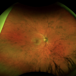
66-year-old white male with Gardner Syndrome (colon resection in 1991), who has two children with Gardner Syndrome, presented to Dr. Zocchi with an RD in the fellow eye that was successfully repaired with a pneumatic retinopexy. Currently 20/20 OU with IOP of 7 OD and 14 OS; no RAPD; PCIOL OU. Dr. Zocchi got oral permission by the patient to have these put into the Retina Image Bank. Although the CHRPE like lesions (2 OD) are not bilateral, we both think these lesions represent "retinal pigment epithelial hamartomas associated with familial adenomatous polyposis (RPEH-FAP)" as Shields described in their Intraocular Tumors book. One lesion is located superiorly and is pigmented with depigmented margins; the temporal lesion is atrophic with minimal remaining pigment hypertrophy.
Photographer: Karin Aletter
Imaging device: Optos CA
Condition/keywords: Gardner Syndrome, RPEH-FAP
-
 Congenital Retinal Pigment Epithelial Hypertrophy (CHRPE) Associated with Gardner's Syndrome
Mar 13 2018 by Olivia Rainey
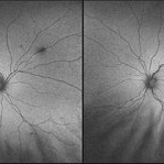
Ultra-wide field fundus autofluorescence images of a 14-year-old patient with congenital retinal pigment epithelial hypertrophy affecting both eyes as a manifestation of Gardner's Syndrome.
Photographer: Olivia Rainey
Imaging device: Optos
Condition/keywords: bilateral, familial adenomatous polyposis, fundus autofluorescence (FAF), Gardner Syndrome, hypofluorescent lesions, Optos, ultra-wide field imaging
-
 Gardner Syndrome - Right
Dec 21 2016 by Tony Tsai, MD, FASRS
9-year-old male with multiple hypertrophic RPE lesions OU characteristic of the kind seen with familial adenomatous polyposis. Genetic testing revealed he was positive for APC mutation. No previously known family history.
Photographer: Reina Hernandez, Retinal Consultants, Sacramento CA
Condition/keywords: Gardner Syndrome
-
 Gardener Syndrome - Left
Dec 21 2016 by Tony Tsai, MD, FASRS
9-year-old male with multiple hypertrophic RPE lesions OU characteristic of the kind seen with familial adenomatous polyposis. Genetic testing revealed he was positive for APC mutation. No previously known family history.
Photographer: Reina Hernandez, Retinal Consultants, Sacramento CA
Condition/keywords: Gardner Syndrome
-
 Bear Tracks
Dec 27 2016 by Elad Moisseiev, MD
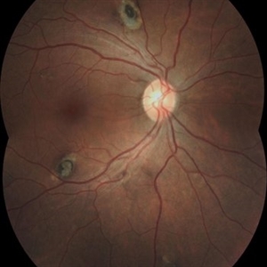
A 34-year-old man with bear tracks noted on routine exam. There was no personal or family history of Gardner syndrome or any GI malignancy.
Photographer: Galit Yair-Pur
Condition/keywords: bear tracks
-
 Congenital Hypertrophy of the Retinal Pigment Epithelium
Nov 11 2019 by Jessica Norkus
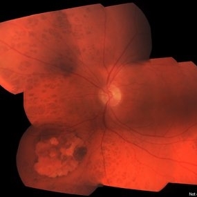
Bilateral Optos ultra wide field imaging of a 31-year-old female patient with CHRPE lesions. Lesions in OD were suspicious of Gardner Syndrome due to familial history of cancerous polyps in colon. Patient underwent colonoscopy and was deemed clear.
Photographer: Jessica Norkus, COA, Retina Specialists of Michigan
Imaging device: Optos Ultra Wide Field Camera
Condition/keywords: bear tracks, bilateral, color fundus photograph, color photo, congenital hypertrophy of the retinal pigment epithelium (CHRPE), fundus autofluorescence (FAF), fundus photograph, lacunae, macula, optic disc, Optos, pseudocolor, ultra-wide field imaging
-
 Benign Lobular Inner Nuclear Layer Proliferations (BLIP)
Apr 15 2024 by Virginia Gebhart
29 year old male with multiple flat CHRPE lesions, genetic testing negative for ACP genes associated with Gardner syndrome. Multiple intraretinal amelanotic lesions consistent with Benign Lobular Inner Nuclear Layer Proliferations (BLIP) of the retina
Photographer: Virginia Gebhart
Imaging device: Topcon
Condition/keywords: BLIP Benign Lobular Inner Nuclear Layer Proliferations, CHRPE, congenital hypertrophy of the retinal pigment epithelium (CHRPE)
-
 Gardner's Syndrome (FAP)
Nov 3 2023 by Virginia Gebhart
43 year-old female with multiple CHRPE lesions consistent with Gardner's Syndrome. Patient had colectomy at age 21. Patient is one of 7 children, 6 had FAP, 3 had colon removal and are alive and well, 3 have passed away from colon cancer. Maternal mother and grandmother also passed away from colon cancer. Patient has 2 children, one son (17) also has Gardner's Syndrome (FAP)
Photographer: Virginia Gebhart
Imaging device: Topcon
Condition/keywords: CHRPE, Gardner Syndrome
-
 Gardner's Syndrome
Nov 10 2023 by Virginia Gebhart
17-year-old male with multiple pigmented spots most likely related to Gardner's Syndrome. Pt has not been diagnosed with FAP at this time, however pt receives regular screenings. Extensive maternal family hx of FAP syndrome and colon cancer. Pt's mother has FAP, who had colon resection. Pt's 2 aunts, 1 uncle, grandmother and great grandmother all passed from colon cancer. Pt has multiple maternal cousins diagnosed with FAP
Photographer: Virginia Gebhart
Imaging device: Topcon
Condition/keywords: CHRPE, congenital hypertrophy of the retinal pigment epithelium (CHRPE), familial adenomatous polyposis, Gardner Syndrome
-
 CHRPE Spots
Jun 13 2025 by Brandon I Fram, MD
21 year-old, no past medical or family histories, with diffuse CHRPE spots suspicious for Gardner syndrome/FAP
Condition/keywords: bear tracks, CHRPE, congenital hypertrophy of the retinal pigment epithelium (CHRPE), familial adenomatous polyposis, Gardner Syndrome
 Loading…