Search results (7 results)
-
 Gardner Syndrome
Dec 12 2018 by John S. King, MD
66-year-old white male with Gardner Syndrome (colon resection in 1991), who has two children with Gardner Syndrome, presented to Dr. Zocchi with an RD in the fellow eye that was successfully repaired with a pneumatic retinopexy. Currently 20/20 OU with IOP of 7 OD and 14 OS; no RAPD; PCIOL OU. Dr. Zocchi got oral permission by the patient to have these put into the Retina Image Bank. Although the CHRPE like lesions (2 OD) are not bilateral, we both think these lesions represent "retinal pigment epithelial hamartomas associated with familial adenomatous polyposis (RPEH-FAP)" as Shields described in their Intraocular Tumors book. One lesion is located superiorly and is pigmented with depigmented margins; the temporal lesion is atrophic with minimal remaining pigment hypertrophy.
Photographer: Karin Aletter
Imaging device: Optos CA
Condition/keywords: Gardner Syndrome, RPEH-FAP
-
 Congenital Retinal Pigment Epithelial Hypertrophy (CHRPE) Associated with Gardner's Syndrome
Mar 13 2018 by Olivia Rainey
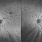
Ultra-wide field fundus autofluorescence images of a 14-year-old patient with congenital retinal pigment epithelial hypertrophy affecting both eyes as a manifestation of Gardner's Syndrome.
Photographer: Olivia Rainey
Imaging device: Optos
Condition/keywords: bilateral, familial adenomatous polyposis, fundus autofluorescence (FAF), Gardner Syndrome, hypofluorescent lesions, Optos, ultra-wide field imaging
-
 Gardner Syndrome - Right
Dec 21 2016 by Tony Tsai, MD, FASRS
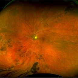
9-year-old male with multiple hypertrophic RPE lesions OU characteristic of the kind seen with familial adenomatous polyposis. Genetic testing revealed he was positive for APC mutation. No previously known family history.
Photographer: Reina Hernandez, Retinal Consultants, Sacramento CA
Condition/keywords: Gardner Syndrome
-
 Bear Tracks
Nov 10 2020 by Ronald Coriasso
Fundus photo of 68-year-old female with history of plaquenil use. Her findings are most consistent with bear tracks, however these kinds of lesions can be indicative of familial adenomatous polyposis (FAP).
Photographer: Ronald Coriasso
Imaging device: OPTOS
Condition/keywords: bear tracks, familial adenomatous polyposis
-
 Gardener Syndrome - Left
Dec 21 2016 by Tony Tsai, MD, FASRS
9-year-old male with multiple hypertrophic RPE lesions OU characteristic of the kind seen with familial adenomatous polyposis. Genetic testing revealed he was positive for APC mutation. No previously known family history.
Photographer: Reina Hernandez, Retinal Consultants, Sacramento CA
Condition/keywords: Gardner Syndrome
-
 Gardner's Syndrome
Nov 10 2023 by Virginia Gebhart
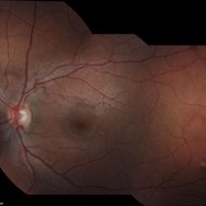
17-year-old male with multiple pigmented spots most likely related to Gardner's Syndrome. Pt has not been diagnosed with FAP at this time, however pt receives regular screenings. Extensive maternal family hx of FAP syndrome and colon cancer. Pt's mother has FAP, who had colon resection. Pt's 2 aunts, 1 uncle, grandmother and great grandmother all passed from colon cancer. Pt has multiple maternal cousins diagnosed with FAP
Photographer: Virginia Gebhart
Imaging device: Topcon
Condition/keywords: CHRPE, congenital hypertrophy of the retinal pigment epithelium (CHRPE), familial adenomatous polyposis, Gardner Syndrome
 Loading…