Search results (21 results)
-
 Reticular Drusen, Doyne's Honeycomb Retinal Dystrophy, Malattia Leventinese, Familial Dominant Drusen
Feb 22 2018 by Nichole Lewis
Reticular Drusen, Doyne's Honeycomb Retinal Dystrophy, Malattia Leventinese, Familial Dominant Drusen
Photographer: Nichole Lewis
Condition/keywords: Doyne's Honeycomb, Familial Dominant Drusen, Malattia Leventinese, reticular drusen
-
 Doyne Honeycomb Retinal Dystrophy
Sep 29 2020 by Navneet Mehrotra, DNB
Left eye fundus photograph of a 36-year-old female with decreased vision both eyes for six months. Father also had a similar retinal disorder.
Photographer: Dr Navneet Mehrotra
Imaging device: TRC- NW8F
Condition/keywords: Doyne's Honeycomb, drusen, Malattia Leventinese
-
 Submacular Hemorrhage Due to Malattia Leventinese
Dec 29 2018 by Darin R. Goldman, MD
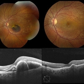
Fundus photograph and OCT of a 37-year-old female with a presumed diagnosis of Malattia Leventinese. One of the more characteristic features of this condition, as seen with this case, is the radiating pattern of drusen and particularly their location nasal to the optic nerve. There is a submacular hemorrhage due to a secondary CNV, which resolved after a series of anti-VEGF therapy with ranibizumab. The followup images (right side) are from 3.5 months after presentation where her VA recovered to 20/20.
Photographer: Robin Lapointe, CRA, Retina Group of Florida
Imaging device: Topcon TRC 50 DX
Condition/keywords: Doyne's Honeycomb, familial drusen, Malattia Leventinese, submacular hemorrhage
-
---thumb.jpg/image-square;max$300,300.ImageHandler) Doyne's
Doyne's
-
---thumb.jpg/image-square;max$300,300.ImageHandler) Drusen
Drusen
-
---thumb.jpg/image-square;max$300,300.ImageHandler) Drusen
Drusen
-
---thumb.jpg/image-square;max$300,300.ImageHandler) Doyne's
Doyne's
-
---thumb.jpg/image-square;max$300,300.ImageHandler) Doyne's
Doyne's
-
---thumb.jpg/image-square;max$300,300.ImageHandler) Drusen
Drusen
-
---thumb.jpg/image-square;max$300,300.ImageHandler) Doyne's
Doyne's
-
---thumb.jpg/image-square;max$300,300.ImageHandler) Doyne's
Doyne's
-
---thumb.jpg/image-square;max$300,300.ImageHandler) Drusen
Drusen
-
---thumb.jpg/image-square;max$300,300.ImageHandler) Drusen
Drusen
-
 Doyne Honeycomb Retinal Dystrophy
Sep 29 2020 by Navneet Mehrotra, DNB
Right eye fundus photograph of a 36-year-old female with decreased vision both eyes for six months. Father also had a similar retinal disorder.
Photographer: Dr Navneet Mehrotra, Retina Care, Ahmedabad
Imaging device: TRC- NW8F
Condition/keywords: Doyne's Honeycomb, familial drusen, Malattia Leventinese
-
 A rare case of a 45-year-old male with choroidal neovascular membrane in Familial Dominant Drusen (Doyne Honeycomb Drusen) in both eyes treated with intravitreal injections.
Nov 30 2022 by SHRADDHA ASHOK CHANDORKAR, DNB DO FVRS
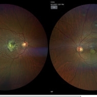
A 45-year-old man presented with diminution of vision in both eyes with metamorphopsia, which was painless and gradually progressive in nature. BCVA at presentation were 6/40 and 6/36 for the right and left eye respectively. Anterior segment examination of both eyes was unremarkable. IOP were within normal limits. Fundus examination showed bilateral numerous yellowish white round closely spaced lesions extending radially from the vascular arcades till the periphery associated with an elevated grayish macular choroidal neovascular membrane (CNV) with multiple drusen in the macular area and posterior pole. Impression was Familial Dominant Drusen (Doyne Honeycomb Drusen) associated with CNVM, both eyes. Color fundus photograph and autofluorescence showed Familial Dominant Drusen with CNVM. Subsequently , the patient underwent periodic intravitreal injections of Ranibizumab in both eyes under guarded visual prognosis, for which he tolerated well.
Photographer: NATIONAL INSTITUTE OF OPHTHALMOLOGY, PUNE
Imaging device: ZEISS CLARUS
Condition/keywords: choroidal neovascular membrane (CNVM), Doyne's Honeycomb, FAMILIAL DOMINANT DRUSEN, IMIM (Online Mendelian Inheritance in Man), intravitreal injection, Malattia Leventinese
-
 Familial Dominant Drusen
Mar 13 2025 by T. P . VIGNESH, MBBS,MS
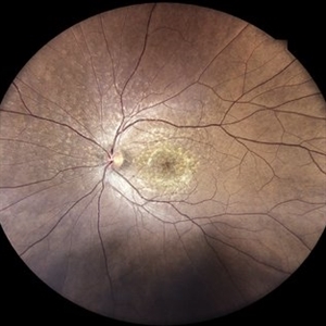
Fundus photograph of an 42-year-old man with familial dominant drusen.
Photographer: Sivanath
Imaging device: EIDON
Condition/keywords: Doyne's Honeycomb
-
 A Crack in the Honeycomb
Jul 28 2025 by Malvika Singh
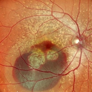
Fundus photograph of a 44 year old with Doyne's Honeycomb Retinal Dystrophy (autosomal dominant drusen) and a subretinal bleed.
Photographer: Dr Malvika Singh, Retina Foundation, Ahmedabad, India
Imaging device: Mirante SLO/OCT
Condition/keywords: Doyne's Honeycomb, retinal dystrophy, subretinal blood
-
 RPE - Rest In Peace (RIP)
Dec 17 2025 by SHRADDHA RAJ SHRIVASTAVA
Right eye pseudocolor fundus photo of a 50 year old patient, known case of bilateral familial dominant drusens with right eye CNVM, having undergone multiple intravitreal anti-VEGF injections. Image shows a CDR of 0.3:1, with numerous drusens at macula with residual lipid exudation from CNVM, along the infero-temporal arcade. Temporal to the fovea, we can see a vertical hyperpigmented line corresponding to retracted and redundant torn Retinal pigment epithelium, leaving behind a well circumscribed area of depigmented fundus with bare Bruch's membrane underlying the retina, findings suggestive of an RPE tear post multiple intravitreal injections.
Photographer: Dr. Shraddha Raj Shrivastava
Imaging device: Nidek Mirante SLO/OCT (Confocal scanning/Spectral domain OCT)
Condition/keywords: choroidal neovascular membrane (CNVM), Doyne's Honeycomb, FAMILIAL DOMINANT DRUSEN, lipid exudation, retinal pigment epithelium, RPE Rip
-
 RPE - Rest In Peace (RIP)
Dec 17 2025 by SHRADDHA RAJ SHRIVASTAVA
Right eye G-FAF photo of a 50 year old patient, known case of bilateral familial dominant drusens with right eye CNVM, having undergone multiple intravitreal anti-VEGF injections. Fundus autofluorescence better highlights the area of RPE tear in right eye (temporal to fovea), which shows hypoautofluorescence due to lack of RPE and its pigments which accounts for autofluorescence signal. Whereas the linear hyperautofluorescence, represents the torn bunched up retinal pigment epithelium.
Photographer: Dr. Shraddha Raj Shrivastava
Imaging device: Nidek Mirante SLO/OCT (Confocal scanning/Spectral domain OCT)
Condition/keywords: choroidal neovascular membrane (CNVM), Doyne's Honeycomb, FAMILIAL DOMINANT DRUSEN, lipid exudation, retinal pigment epithelium, RPE Rip
-
 RPE - Rest In Peace (RIP)
Dec 17 2025 by SHRADDHA RAJ SHRIVASTAVA
Right eye RETRO mode fundus image of a 50 year old patient, known case of bilateral familial dominant drusens with right eye CNVM, having undergone multiple intravitreal anti-VEGF injections. Among other findings, this novel imaging technique highlights the presence of an extrafoveal RPE tear - post multiple intravitreal injections.
Photographer: Dr. Shraddha Raj Shrivastava
Imaging device: Nidek Mirante SLO/OCT (Confocal scanning/Spectral domain OCT)
Condition/keywords: choroidal neovascular membrane (CNVM), Doyne's Honeycomb, FAMILIAL DOMINANT DRUSEN, lipid exudation, retinal pigment epithelium, RPE Rip
 Loading…