Search results (52 results)
-
---thumb.jpg/image-square;max$300,300.ImageHandler) Coats Disease
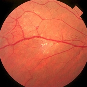
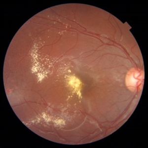
Coats Disease
Oct 30 2012 by Lihteh Wu, MD
Fundus photograph of a 29-year-old man with no significant medical or ocular history. Patient complained of progressive loss of vision over the past few months. Notice the lipid exudation over the macula and the hyperplastic RPE.
Condition/keywords: hyperplastic retinal pigment epithelium (RPE), lipid exudation, retinal pigment epithelium
-
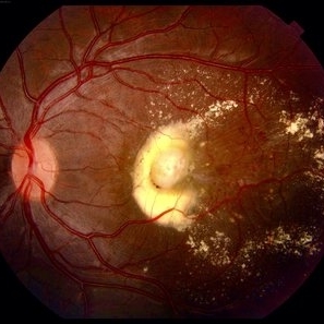
 Coats Disease
Oct 9 2012 by Alan D. Letson, MD
Coats Disease
Photographer: Beverly Radcliffe
Condition/keywords: retinal macroaneurysm, retinal telangiectasia
-
 Coats Disease
Apr 16 2015 by Rita Couceiro, MD, MS
Fundus photograph and fluorescein angiography pictures of a 13-year-old girl with Coats Disease, showing abnormal telangiectatic vessels and intense exsudation in the inferior retinal periphery of the left eye.
Condition/keywords: Coats' disease, retinal telangiectasia
-
 Coats Disease
Feb 7 2013 by Raj K. Maturi, MD
8-year-old male, flourescein angiogram, venous phrase, OD.
Photographer: Stephan Morrow, Midwest Eye Institute Indianapolis Indiana
Imaging device: Heidelberg Spectralis
Condition/keywords: Spectralis
-
---thumb.jpg/image-square;max$300,300.ImageHandler) Coats Disease
Coats Disease
Oct 30 2012 by Lihteh Wu, MD
FA frame showing blocked fluorescence from the massive lipid exudation. There is also hyperfluorescence secondary to vascular leakage and hypofluorescence from the hyperplastic RPE. Superotemporal to the fovea there are areas of telangiectasia.
Condition/keywords: massive lipid exudation, retinal pigment epithelium, retinal telangiectasia
-
---thumb.JPG/image-square;max$300,300.ImageHandler) Coats Disease
Coats Disease
Oct 11 2012 by Anat Loewenstein, MD
Fluorescein angiography of 6 -year-old girl whose parents have noticed leukocoria in her right eye. On examination severe exudative retinal detachment was diagnosed. On FA of the right eye peripehral capillary non perfusion and peripheral capillary dilatations were seen.
Photographer: Galit Yair-Pur
-
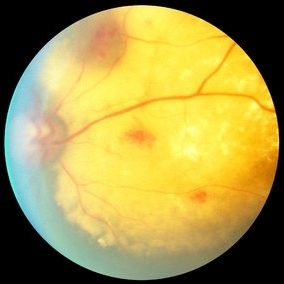
 Chorioretinal Scar
May 16 2017 by Olivia Rainey
Fundus photograph of an 17-year-old male with a macular scar affecting his right eye secondary to exudation from Coats disease.
Photographer: Olivia Rainey
Imaging device: Topcon 50dx
Condition/keywords: 20 degrees, chorioretinal scar, Coats' disease, color fundus photograph, color photo, fundus photograph
-
---thumb.jpg/image-square;max$300,300.ImageHandler) Coats disease
Coats disease
Jan 11 2013 by Hyung-Woo Kwak, MD
Fundus imaging shows hemorrhage and hard exudates from leaking blood vessel.
Photographer: Taegi Kim, Kyung Hee Univsersity Hospital, Seoul
Imaging device: Zeiss f 450 plus
Condition/keywords: argon photocoagulation
-
 Coats Disease
Jul 7 2022 by Gabriel Costa Andrade, PhD
Fundus photograph of a 31-year-old man with no medical or ocular history. Patient complained of progressive loss of vision over the past few months OS. Notice the lipid exudation over the macula and telangiectatic vessels.
Photographer: Dr Gabriel Andrade
Condition/keywords: Coats' disease
-
 Coats Disease
Feb 7 2013 by Raj K. Maturi, MD
8-year-old male, Heidelberg Spectralis OCT, OD.
Photographer: Stephan Morrow, Midwest Eye Institute Indianapolis Indiana
Imaging device: Heidelberg Spectralis
Condition/keywords: optical coherence tomography (OCT), Spectralis
-
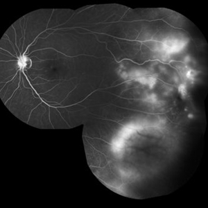
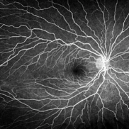
 Coats Disease
Sep 13 2013 by Maria Ana Martinez-Castellanos, MD
Peripheral fundus angiogram in a 2-years-old boy with Coat's disease.
Photographer: Maria A. Martinez-Castellanos. Asociacion para Evitar la Ceguera en Mexico
Imaging device: RetCAm II
Condition/keywords: pediatic retina, serous retinal detachment, vascular anomaly, vascular occlusions
-
 Coats Disease Slide 1
Oct 22 2012 by Ronald C. Gentile, MD
A unilateral, sub-retinal, and yellowish exudative lesion with associated retinal telangiectasias involving the nasal retina. Refractile elements can be seen and represent cholesterol crystals.
Photographer: The New York Eye & Ear Infirmary Department of Medical Imaging
Condition/keywords: congenital retinal telangiectasis
-
 Coats Disease
May 27 2016 by Olivia Rainey
Composite fluorescein angiogram of the left eye of a man with Coats Disease.
Photographer: Olivia Rainey
Imaging device: Heidelberg Spectralis
Condition/keywords: Coats' disease, composite, fluorescein angiogram (FA), fluorescein leakage, Heidelburg Spectralis
-
 Serous Retinal Detachment in Coats Disease
Mar 31 2014 by Maria Ana Martinez-Castellanos, MD
Fundus photograph of a 3-year-old boy with low vision, esotropia and leukocoria.
Photographer: Maria A. Martinez-Castellanos. Asociacion para Evitar la Ceguera en Mexico
Imaging device: RetCam II
Condition/keywords: pediatic retina, vascular anomaly
-
 Coats Disease
Feb 7 2013 by Raj K. Maturi, MD
8-year-old male, flourescein angiogram, late phrase 4:22 minutes, OD.
Photographer: Stephan Morrow, Midwest Eye Institute Indianapolis Indiana
Imaging device: Heidelberg Spectralis
Condition/keywords: Spectralis
-
 Coats Disease Slide 2
Oct 22 2012 by Ronald C. Gentile, MD
Telangiectatic vessels are seen above the subretinal exudation. The abnormal vessels are tortuous and dilated.
Photographer: The New York Eye & Ear Infirmary Department of Medical Imaging
Condition/keywords: congenital retinal telangiectasis
-
---thumb.jpg/image-square;max$300,300.ImageHandler) Coats Disease
Coats Disease
Oct 30 2012 by Lihteh Wu, MD
FA frame showing peripheral telangiectasia and some vascular leakage.
Condition/keywords: peripheral telangiectasia
-
 Coats Disease Slide 3
Oct 22 2012 by Ronald C. Gentile, MD
Flourescein angiogram with early filling of telangectatic vessels.
Photographer: The New York Eye & Ear Infirmary Department of Medical Imaging
Condition/keywords: congenital retinal telangiectasis
-
 Coats Disease Slide 4
Oct 22 2012 by Ronald C. Gentile, MD
Flourescein angiogram with progressive filling of telangectatic vessels. The abnormal vessels become more prominent and the dilated aneurysmal-like vessels fill.
Photographer: The New York Eye & Ear Infirmary Department of Medical Imaging
Condition/keywords: congenital retinal telangiectasis
-
 Coats Disease Slide 5
Oct 22 2012 by Ronald C. Gentile, MD
Flourescein angiogram with leakage of the telangectatic and dilated aneurysmal-like abnormal vessels.
Photographer: The New York Eye & Ear Infirmary Department of Medical Imaging
Condition/keywords: congenital retinal telangiectasis
-
 Coats Disease Slide 6
Oct 22 2012 by Ronald C. Gentile, MD
Late flourescein angiogram with progressive leakage of of the telangectatic vessels and staining of the entire lesion.
Photographer: The New York Eye & Ear Infirmary Department of Medical Imaging
Condition/keywords: congenital retinal telangiectasis
-
 Coats' Disease
Mar 4 2017 by Hashim Ali Khan, OD, FAAO
Color Fundus Image of an 18-year-old girl with Coats disease.
Condition/keywords: Coats' disease, exudates over the posterior pole, macular edema, macular telangiectasia
-
 Coats' Disease
Sep 2 2025 by Drew Mitchell
Optos color photograph of a young boy with Coats disease. Extensive subretinal exudation that is encroaching towards macula. There are peripheral berry aneurysms with localized area of subretinal fluid. Discussed treatment options including laser photocoagulation of aneurysms. Risks benefits and alternatives discussed including possible need for cryo.
Photographer: Drew Mitchell, OCT-C
Imaging device: Optos California
Condition/keywords: Coats' disease
-
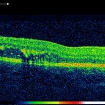 OCT of 18-Year-Old Girl With Coats Disease
Mar 4 2017 by Hashim Ali Khan, OD, FAAO
OCT B scan showing intraretinal exudation and fluid.
Condition/keywords: B scan ultrasound, Coats' disease, exudate
-
 Fluorescein Angiography of Patient with Coat's Disease.
Oct 20 2020 by Anfisa Ayalon, MD
Fundus fluorescein angiography of 35-year-old female with right eye asymptomatic coats disease.
Photographer: Anfisa Ayalon, MD., Meir Medical Center, Kfar Saba, Israel.
Imaging device: California, Optos 200 DTX
Condition/keywords: Coats' disease, fluorescein leakage, leakage
 Loading…