Search results (48 results)
-
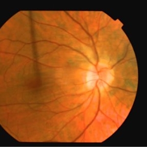
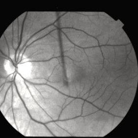
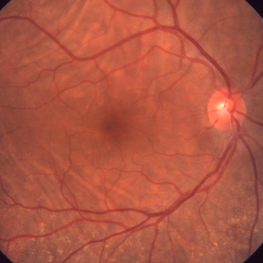 Bilateral Idiopathic Choroidal Folds
Jan 11 2013 by Gerardo Garcia-Aguirre, MD
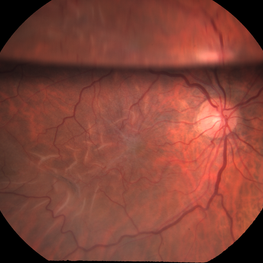
Fundus photograph of the right eye showing choroidal folds.
Imaging device: Zeiss FF4
Condition/keywords: choroidal folds
-
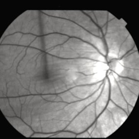
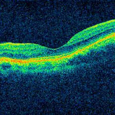 Bilateral Idiopathic Choroidal Folds - OCT
Jan 11 2013 by Gerardo Garcia-Aguirre, MD
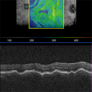
OCT of the macula showing choroidal folds.
Photographer: Gerardo Garcia-Aguirre, MD
Imaging device: Zeiss Cirrus HD OCT
Condition/keywords: choroidal folds
-
 Choroidal folds due to hypotony
Jan 11 2013 by Alex P. Hunyor, MD
Choroidal folds due to hypotony
Condition/keywords: choroidal folds, hypotonous retinopathy
-
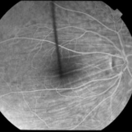
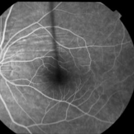
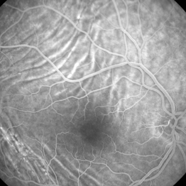 Idiopathic Choroidal Folds - Fluorescein Angiogram
Jan 11 2013 by Gerardo Garcia-Aguirre, MD
Fluorescein Angiogram showing choroidal folds.
Imaging device: Zeiss FF4
Condition/keywords: choroidal folds
-
 Choroidal Folds in NVAMD
Sep 10 2012 by James B. Soque, CRA, OCT-C, COA, FOPS
75 y/o Female, FA with Superior and Inferior Choroidal Folds, and Neovascular Age Related Macular Degeneration of the Right Eye.
Photographer: James B Soque, CRA, COA
Imaging device: TRC-50DX
Condition/keywords: choroidal folds
-
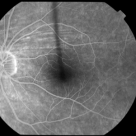
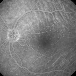 Choroidal Folds - Fluorescein Angiogram
Jan 11 2013 by Gerardo Garcia-Aguirre, MD
Fluorescein angiogram.
Photographer: Gerardo Garcia-Aguirre, MD
Imaging device: Zeiss FF4
Condition/keywords: choroidal folds
-
 Post Choroidal Folds OD
Mar 12 2014 by Manish Nagpal, MD, FRCS (UK), FASRS
32-year-old male had presented with extensive choroidal folds. Oral and sub tenon steroids resolved the folds and only a few residual stria are seen with good visual recovery.
Photographer: Pooja Barot
Condition/keywords: choroidal folds
-
 Choroidal Folds
Nov 28 2014 by Thomas A. Ciulla, MD, MBA, FASRS
This 53-year-old man was noted to have choroidal folds right greater than left. The visual acuity was normal at 20/15. The choroidal folds are visible on OCT, especially on the vertical cuts that image across the horizontal folds. Angiography revealed staining of the folds without CNVM, choroidal mass, or optic nerve edema.
Photographer: Charlotte Harris
Condition/keywords: bilateral chorioretinal folds, choroidal folds
-
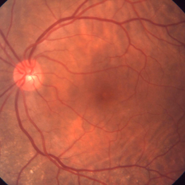 Bilateral idiopathic choroidal folds
Jan 11 2013 by Gerardo Garcia-Aguirre, MD
Fundus photograph showing choroidal folds.
Imaging device: Zeiss ff4
Condition/keywords: choroidal folds
-
 Choroidal Folds
-
 PRE CF OD June 5, 2013
Mar 12 2014 by Manish Nagpal, MD, FRCS (UK), FASRS
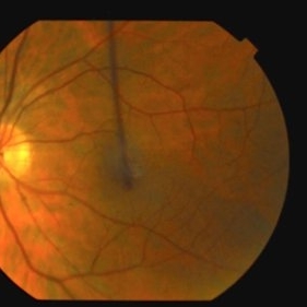
Fundus photo of a 32-year-old male presenting with post traumatic choroidal folds and hypotony.
Photographer: Pooja Barot
Condition/keywords: choroidal folds
-
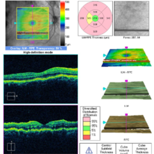
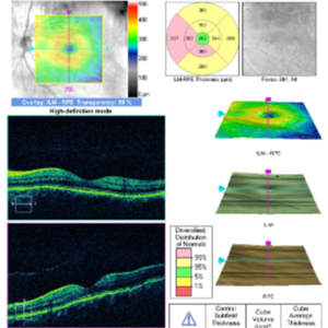
 Bilateral Idiopathic Choroidal Folds
Jan 11 2013 by Gerardo Garcia-Aguirre, MD
3D reconstruction of the RPE showing choroidal folds.
Photographer: Gerardo Garcia-Aguirre, MD
Imaging device: Zeiss Cirrus HD OCT
Condition/keywords: choroidal folds
-
 Choroidal Folds
Nov 28 2014 by Thomas A. Ciulla, MD, MBA, FASRS
This 53-year-old man was noted to have choroidal folds right greater than left. The visual acuity was normal at 20/15. The choroidal folds are visible on OCT, especially on the vertical cuts that image across the horizontal folds. Angiography revealed staining of the folds without CNVM, choroidal mass, or optic nerve edema.
Photographer: Charlotte Harris
Condition/keywords: bilateral chorioretinal folds, choroidal folds
-
 Choroidal Folds
Nov 28 2014 by Thomas A. Ciulla, MD, MBA, FASRS
This 53 -year-old man was noted to have choroidal folds right greater than left. The visual acuity was normal at 20/15. The choroidal folds are visible on OCT, especially on the vertical cuts that image across the horizontal folds. Angiography revealed staining of the folds without CNVM, choroidal mass, or optic nerve edema.
Condition/keywords: bilateral chorioretinal folds, choroidal folds
-
 Hypotony Maculopathy
Apr 1 2019 by Anfisa Ayalon, MD
Fundus autofluorescence image of 81-year-old male with right eye ocular hypotony due to leaking bleb. Note severe hypotony maculopathy, peripheral choroidal detachments, multiple chorioretinal folds.
Photographer: Anfisa Ayalon, MD., Meir Medical Center, Kfar Saba, Israel.
Imaging device: California, Optos 200 DTX
Condition/keywords: choroidal detachment, choroidal folds, fundus autofluorescence (FAF), hypotonous retinopathy, hypotony maculopathy
-
 Central Serous Chorioretinopathy (CSCR) With Choroidal Folds
Jun 2 2015 by Mallika Goyal, MD
Right fundus photograph of a 56-year-old male with CSCR with a hypopigmented spot corresponding to the FFA leak with choroidal folds (seen on OCT); subretinal fluid was limited to an area superior to macular centre, and patient presented with complaint of inferior field loss for 4 weeks.
Photographer: Mallika Goyal, MD, Apollo Health City, Jubilee Hills, Hyderabad
Condition/keywords: central serous chorioretinopathy (CSCR)
-
 Epiretinal Membrane
Mar 3 2017 by Nichole Lewis
64-year-old female in a post op period with an epiretinal membrane and folds with a gas bubble. S/P repaired partial retinal detachment with multiple tears.
Photographer: Nichole Lewis
Condition/keywords: choroidal folds, epiretinal membrane (ERM)
-
 Choroidal Folds
Nov 28 2014 by Thomas A. Ciulla, MD, MBA, FASRS
This 53-year-old man was noted to have choroidal folds right greater than left. The visual acuity was normal at 20/15. The choroidal folds are visible on OCT, especially on the vertical cuts that image across the horizontal folds. Angiography revealed staining of the folds without CNVM, choroidal mass, or optic nerve edema.
Photographer: Charlotte Harris
Condition/keywords: bilateral chorioretinal folds, choroidal folds
-
 Choroidal Folds
-
 Choroidal Folds
Nov 28 2014 by Thomas A. Ciulla, MD, MBA, FASRS
This 53-year-old man was noted to have choroidal folds right greater than left. The visual acuity was normal at 20/15. The choroidal folds are visible on OCT, especially on the vertical cuts that image across the horizontal folds. Angiography revealed staining of the folds without CNVM, choroidal mass, or optic nerve edema.
Condition/keywords: bilateral chorioretinal folds, choroidal folds
-
 Choroidal Folds
Nov 28 2014 by Thomas A. Ciulla, MD, MBA, FASRS
This 53-year-old man was noted to have choroidal folds right greater than left. The visual acuity was normal at 20/15. The choroidal folds are visible on OCT, especially on the vertical cuts that image across the horizontal folds. Angiography revealed staining of the folds without CNVM, choroidal mass, or optic nerve edema.
Photographer: Charlotte Harris
Condition/keywords: bilateral chorioretinal folds, choroidal folds
-
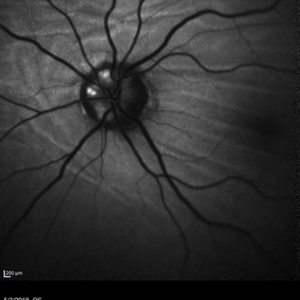 Choroidal Folds and Optic Disc Drusen
Aug 1 2018 by Emily Cooper
Fundus autofluorescence photo of a 62-year-old man who presented for evaluation of choroidal folds and optic disc drusen. He is currently following up with neuro-ophthalmology and has suspected intracranial hypertension.
Photographer: Emily Cooper, Retina Specialists of Michigan
Condition/keywords: choroidal folds, drusen of optic disc
-
 Choroidal Folds
Nov 28 2014 by Thomas A. Ciulla, MD, MBA, FASRS
This 53-year-old man was noted to have choroidal folds right greater than left. The visual acuity was normal at 20/15. The choroidal folds are visible on OCT, especially on the vertical cuts that image across the horizontal folds. Angiography revealed staining of the folds without CNVM, choroidal mass, or optic nerve edema.
Photographer: Charlotte Harris
Condition/keywords: bilateral chorioretinal folds, choroidal folds
-
 Choroidal Folds
Nov 28 2014 by Thomas A. Ciulla, MD, MBA, FASRS
This 53-year-old man was noted to have choroidal folds right greater than left. The visual acuity was normal at 20/15. The choroidal folds are visible on OCT, especially on the vertical cuts that image across the horizontal folds. Angiography revealed staining of the folds without CNVM, choroidal mass, or optic nerve edema.
Photographer: Charlotte Harris
Condition/keywords: bilateral chorioretinal folds, choroidal folds
 Loading…