Search results (206 results)
-
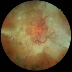
 Tractional Retinal Detachment
Dec 4 2019 by Janet Brazil
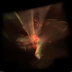
Fundus photograph of a 32-year-old female with severe end-stage diabetic tractional retinal detachment.
Photographer: Janet Atkinson, Eye Associates of New Mexico, Albuquerque, NM
Imaging device: Topcon TRC- 50EX
Condition/keywords: diabetes, proliferative diabetic retinopathy (PDR), tractional retinal detachment
-
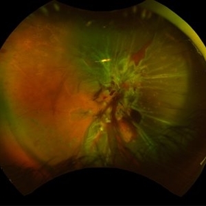
 Sickle Cell Retinopathy
Nov 5 2022 by Mateus Queiroz Corrêa, MD
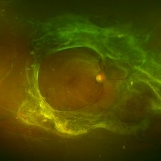
19 -year-old young man with combined rhegmatogenous and tractional retinal detachment secondary to a proliferative sickle retinopathy ( stage V)
Photographer: Mateus Corrêa, Sorocaba Eye Bank Hospital
Imaging device: Optos California
Condition/keywords: Retinal detachment, sickle cell retinopathy
-
 Tractional Retinal Detachment
Sep 27 2012 by Virgilio Morales-Canton, MD
OCT image of a 42-year-old male patient with a localized traction of the superior macula secondary to proliferative diabetic retinopathy.
Imaging device: Cirrus
Condition/keywords: tractional retinal detachment
-
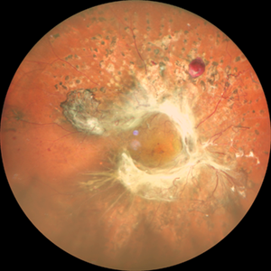
 Diabetic traction retinal detachment
Jan 9 2023 by JORGE SOBERANES
Proliferative diabetic retinopathy with extensive traction retinal detachment in a patient with type 1 diabetes mellitus.
Photographer: Dr. Jorge I. Soberanes, Asociación para Evitar la Ceguera en México.
Imaging device: Zeiss Clarus 700
Condition/keywords: Retinal Detachment, tractional retinal detachment
-
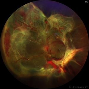
 Displaced & folded macula
Oct 10 2022 by Ricardo Leitão Guerra
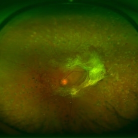
Tractional retinal detachment due to sickle cell retinopathy leading to a displaced and folded appearance of the macula in this 36-yo male. Subretinal bands are also noticed crossing the macula towards inferior retinal detachment area.
Photographer: Ricardo Leitão Guerra
Imaging device: Clarus 700 - Zeiss
Condition/keywords: folds, sickle cell retinopathy, subretinal bands, tractional retinal detachment
-
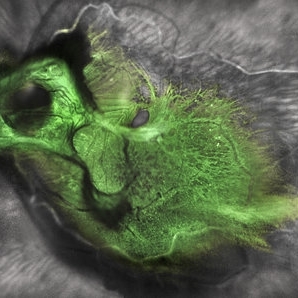 Green Goblin Detachment
Jan 13 2022 by Netan Choudhry, MD, FRCS(C) FASRS
Tractional retinal detachment with macular hole in a 76-year-old female.
Photographer: John Golding BA, Vitreous Retina Macula Specialists of Toronto, OCTane Imaging Lab
Imaging device: Multicolor fundus photo taken on the Spectralis OCT2 (Heidelberg Engineering GmbH).
Condition/keywords: macular hole, Multispectral imaging, tractional retinal detachment
-
 Tractional Detachment of Retina
Aug 21 2024 by Jordyn Beckman
18 year old male with tractional detachment of Retina, chronic macular hole and silicone oil s/p RD repair x2. BCVA CF @2 ft, fellow eye prosthetic.
Photographer: Jordyn Beckman
Imaging device: Optos California
Condition/keywords: Macular hole, preretinal fibrosis, Retinal Detachment, scleral buckle, silicone oil, TRACTION, tractional retinal detachment
-
 Annular Tractional Retinal Detachment
Jul 4 2024 by Hector Gabriel Moreno Solano, MD, MHA
52-year-old Hispanic female patient with a diagnosis of type II diabetes mellitus of 15 years of evolution, comes to the retina service for progressive visual loss in the right eye (single functional eye) with visual acuity of 20/100, Fundus examination reveals laser-modified proliferative diabetic retinopathy with activity + annular tractional retinal detachment with macular involvement.
Photographer: Hector Gabriel Moreno Solano, MD, MHA, HGZ #20 IMSS Puebla.
Imaging device: Mirante
Condition/keywords: macular detachment, proliferative diabetic retinopathy (PDR), tractional retinal detachment
-
000---thumb.jpg/image-square;max$300,300.ImageHandler) Anterior Segment Photo of Emulsified Silicone Oil
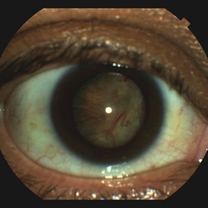
Anterior Segment Photo of Emulsified Silicone Oil
Dec 25 2013 by Dong Yoon Kim, MD
47-year-old woman underwent vitrectomy and silicone oil tampoande for tractional retinal detachment due to proliferative diabetic retinopathy. 8 months after silicone oil tamponade, silicone oil was emulsified. And emulsified silicone oil was observed at anterior chamber.
Condition/keywords: silicone oil, tractional retinal detachment
-
 Combined Traction and Rhegmatogenous Retinal Detachment From Proliferative Diabetic Retinopathy
Mar 27 2025 by Nikhil K Bommakanti, MD
A middle-aged patient presented with a combined traction and rhegmatogenous retinal detachment.
Condition/keywords: Active PDR Tractional retinal Detachment, PDR, Retinal Detachment, rrd, TRD
-
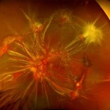
 Diabetic Spider Web
Nov 5 2021 by Joana Roque
59 year-old poorly controlled diabetic patient with proliferative retinopathy and tractional retinal detachment.
Photographer: Joana Roque
Condition/keywords: proliferative diabetic retinopathy (PDR)
-
 Diabetic Traction Retinal Detachment
Mar 21 2013 by Yusuke Oshima, MD, PhD
Wide-field fundus photograph of a 56-year-old man with tractional retinal detachment due to proliferative diabetic retinopathy.
Photographer: Yusuke Takada, Osaka University Graduate School of Medicine
Imaging device: OPTOS 200Tx
-
 Diabetic Tractional Retinal Detachment
May 19 2014 by John W. Kitchens, MD
Severe PDR with non perfusion and a tractional retinal detachment.
Photographer: Ed Slade
Imaging device: Optos 200Tx
Condition/keywords: diabetes
-
 Diabetic Tractional Retinal Detachment
Jan 23 2019 by Olivia Rainey
Ultra-wide field pseudocolor image of an 43-year-old female with a diabetic tractional retinal detachment and a vitreous hemorrhage affecting her right eye.
Photographer: Olivia Rainey
Imaging device: Optos
Condition/keywords: diabetes, diabetic traction detachment, Optos, pan-retinal photocoagulation (PRP), proliferative diabetic retinopathy (PDR), pseudocolor, ultra-wide field imaging, vitreous hemorrhage
-
 Diabetic Tractional Retinal Detachment Involving the Macula OD
Feb 21 2025 by Kaitlyn Anderson
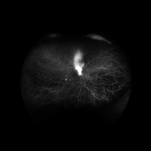
57-year-old female. Diabetic Tractional Retinal Detachment involving the Macula OD. Active Proliferative Diabetic Retinopathy
Photographer: Kaitlyn Anderson TN Retina Nashville TN
Imaging device: Optos Fluorescein Angiogram
Condition/keywords: Active PDR Tractional retinal Detachment
-
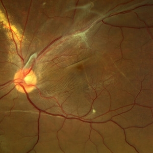
 Macular Hole Due to Proliferative Diabetic Retinopathy
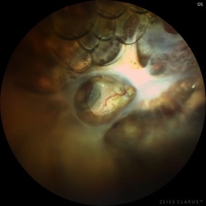
Aug 13 2025 by Ricardo Leitão Guerra
A macular hole formation after anti-VEGF injection prior to vitrectomy for tractional retinal detachment in a patient presenting proliferative diabetic retinopathy.
Photographer: Ricardo Leitão Guerra
Imaging device: ZEISS CLARUS 700
Condition/keywords: macular hole, proliferative diabetic retinopathy (PDR)
-
 Proliferative Diabetic Retinopathy
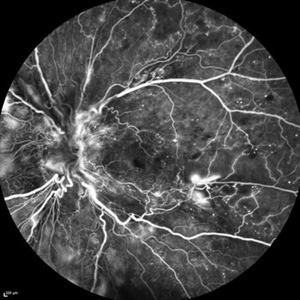
Mar 16 2015 by Matt Poe, COA
IVFA of 53-year-old male with Proliferative Diabetic Retinopathy, Diabetic macular edema, and a tractional retinal detachment.
Photographer: Matt Poe, COA. Northwest Arkansas Retina Associates, Springdale, AR.
Imaging device: Heidelberg HRA
Condition/keywords: neovascularization (NV), proliferative diabetic retinopathy (PDR)
-
 Proliferative Diabetic Retinopathy
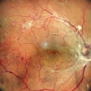
May 24 2024 by Anjana Mirajkar, MS Ophthalmology
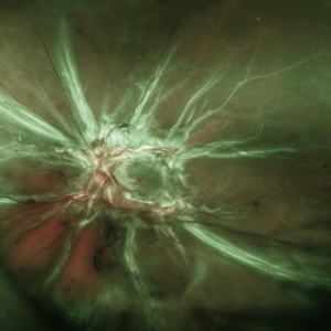
A central photo of a 65 year old female of right eye showing fibro vascular proliferation with neovascularization elsewhere in a case of proliferative diabetic retinopathy.
Photographer: Dr. Anjana Mirajkar -Retina Foundation, Ahmedabad
Imaging device: Mirante-Nidek
Condition/keywords: NVE, proliferative diabetic retinopathy (PDR), tractional retinal detachment
-
---thumb.jpg/image-square;max$300,300.ImageHandler) Proliferative Diabetic Retinopathy (PDR) & Traction Retinal Detachment
Proliferative Diabetic Retinopathy (PDR) & Traction Retinal Detachment
Feb 13 2013 by From the Collections of Thomas M. Aaberg, MD and Thomas M. Aaberg Jr., MD
Florid NV with early macular TRD.
Condition/keywords: neovascularization (NV), tractional retinal detachment
-
 RETINAL DETACHMENT WITH EMULSIFIED SILICONE
Sep 22 2023 by BENITO VERGARA, MD
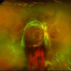
Fundus photograph of a 67-year-old man, with surgical history of phacoemulsification and vitrectomy, in which we can observe a tractional retinal detachment with emulsified silicone oil that precludes the visualization of posterior pole structures.
Photographer: Benito Vergara-Flores, Asociación para Evitar la Ceguera en México
Imaging device: Clarus 700
Condition/keywords: emulsification, tractional retinal detachment
-
 Total Rhegmatogenous and Tractional Retinal Detachment Following Choroidal Melanoma Laser Ablation Treatment
Sep 22 2020 by Sophia El Hamichi, MD
A 69-year-old female, with a history of choroidal melanoma in her left eye with exudative detachment, underwent tumor laser ablation. She then developed a complex combined tractional and rhegmatogenous retinal detachment with a giant retinal tear.
Photographer: Belinda Rodriguez, Murray Ocular Oncology and Retina, Miami
Condition/keywords: tractional retinal detachment
-
 Tractional and Rhegmatogenous Retinal Detachment
Dec 30 2016 by Manish Nagpal, MD, FRCS (UK), FASRS
Combined tractional and rhegmatogenous retinal detachment seen in a case of vasculitis. Two small breaks are noted in the extreme nasal area next to an area of proliferation.
Photographer: Avijit Vishnoi
Condition/keywords: tractional retinal detachment, vasculitis
-
 Tractional Retinal Detachment
Jan 23 2018 by Nilesh K Kanjani, MD
Fundus Photograph of 42-year-old female patient with uncontrolled diabetes show tractional retinal detachment involving macula.
Photographer: Nilesh K Kanjani, Dr Agarwal Eye Hospital, Ahmedabad, India
Condition/keywords: tractional retinal detachment
-
 Tractional Retinal Detachment
Feb 9 2015 by Matt Poe, COA
This patient presented with bilateral tractional retinal detachments secondary to her proliferative diabetic retinopathy. Surprisingly the patient had 20/60 in that eye.
Photographer: Matt Poe, COA. Northwest Arkansas Retina Associates, Springdale, AR.
Condition/keywords: diabetic mellitus, proliferative diabetic retinopathy (PDR), tractional retinal detachment
-
 Tractional Retinal Detachment
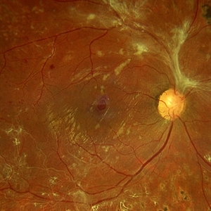
Apr 20 2024 by Tejaswita Verma
Fundus photograph of the right eye of a 62 year old female with tractional retinal detachment in a case of lasered proliferative diabetic retinopathy showing neovascularisation at disc and elsewhere
Photographer: DR. TEJASWITA VERMA
Condition/keywords: Neovascularisation at the Disc (NVD), Neovascularisation elsewhere (NVE), proliferative diabetic retinopathy (PDR)
 Loading…