Search results (24 results)
-
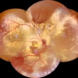 Coats' Disease Montage
Feb 5 2021 by Akansha Sharma
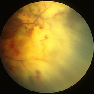
Fundus photograph of a 5-year-old male child who presented with unilateral diminution of vision since one month.
Photographer: Dr. Nivesh Gupta, M.S., Retina Foundation, Ahmedabad
Condition/keywords: angiomatosis retinae, Coats' disease, exudative detachment, subretinal exudates
-
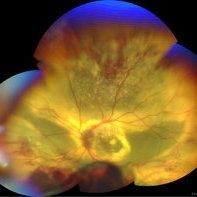 Coats' disease
Sep 7 2022 by Niloofar Piri, MD
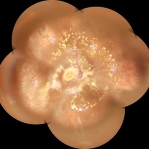
Total exudative RD with extensive subretinal exudates and peripheral telangiectatic vascular anomalies in stage 4 Coats's disease. Patient is a 12 yo who presented with severe eye pain and neovascular glaucoma secondary to the above.
Photographer: Jacob Grodsky, MD
Condition/keywords: Coats' disease
-
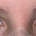 Xanthocoria in Coats' disease
Sep 7 2022 by Niloofar Piri, MD
Xanthocoria secondary to total exudative RD in Coats' disease. Yellow color is secondary to extensive subretinal exudates.
Photographer: Jacob Grodsky, MD
Condition/keywords: Coats' disease, Xanthocoria
-
 Bartonella Posterior Granulomatous Mass with Exudative Detachment Post 2 Months
Sep 26 2020 by Swati Agarwal-Sinha, MD, FASRS
Color fundus photo of the left eye two months post-treatment showing resolved exudative detachment, fibrosed granuloma at the optic disc, tractional detachment at the posterior pole and extensive diffuse subretinal exudates extending all around up to the retinal periphery. No retinal vascular changes are noted.
Photographer: Harry Rosa, University of Florida
Condition/keywords: Bartonella bacteria, exudative detachment
-
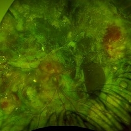 Advanced coats disease
Dec 27 2023 by NIDHI PANWAR, MD FNB FICO
Fundus photograph of 6 year old otherwise healthy boy presented with right eye esotropia and poor vision with fundus picture depicting advanced exudative retinal disease suggestive of coats disease
Photographer: Nidhi Panwar, NMC Royal hospital, Sharjah , UAE
Condition/keywords: Coats disease, subretinal exudates
-
 Coat's Disease
Mar 29 2013 by Henry J. Kaplan, MD
Typical peripheral telangiectatic vessles and subretinal exudates.
Condition/keywords: Coats' disease
-
 Coat's Disease - Widefield Montage
Nov 22 2019 by Gayathri Mohan
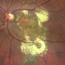
Widefield color fundus photo montage of a patient with RE Coats disease, showing diffuse exudates along with sub foveal nodule.
Photographer: Dr. Gayathri Mohan- Retina Foundation
Imaging device: Mirante, Nidek
Condition/keywords: Coats' disease, subretinal exudates
-
 Coat's Disease with Exudative RD
Feb 12 2025 by Tejaswita Verma
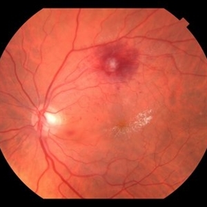
Fundus photo of a 7 year old boy with vision Counting fingers close to face in the right eye and intermittent outward deviation of the right eye observed by parents. Fundus examination shows subretinal exudates, telengiectatic vessels in superotemporal quadrant, intraretinal hemorrhages, macular scar, NVD.
Photographer: DR. TEJASWITA VERMA
Imaging device: MIRANTE
Condition/keywords: Coats' disease, exudative retinal detachment
-
 Coats' Disease with NVI
Sep 18 2020 by Neha Goel, MS DNB FRCS (Glasg)
Slit lamp photograph of the left eye of a 20-year-old male showing NVI and ectropion uveae.
Condition/keywords: bullous retinal detachment, Coats' disease, exudative retinal detachment, neovascularization of iris (NVI), subretinal exudates, telangiectatic vessels
-
 Coats' Disease with NVI
Sep 18 2020 by Neha Goel, MS DNB FRCS (Glasg)
Slit lamp photograph of the left eye of a 20-year-old male showing high bulls retinal detachment with the retina near the posterior capsule of the lens. Presence of telangiectatic vessels points towards Coats' disease.
Condition/keywords: bullous retinal detachment, Coats' disease, exudative retinal detachment, neovascularization of iris (NVI), subretinal exudates, telangiectatic vessels
-
 Coats' Disease with NVI
Sep 18 2020 by Neha Goel, MS DNB FRCS (Glasg)
Montage funds photograph of the left eye of a 20-year-old male showing retinal detachment with sub retinal exudates superiorly and at the macula.
Condition/keywords: bullous retinal detachment, Coats' disease, exudative retinal detachment, neovascularization of iris (NVI), subretinal exudates, telangiectatic vessels
-
 Coats' Disease with NVI
Sep 18 2020 by Neha Goel, MS DNB FRCS (Glasg)
Ultrasound B scan of a 20-year-old male showing exudative retinal detachment.
Condition/keywords: bullous retinal detachment, Coats' disease, exudative retinal detachment, neovascularization of iris (NVI), subretinal exudates, telangiectatic vessels
-
---thumb.JPG/image-square;max$300,300.ImageHandler) Dengue Retinitis
Dengue Retinitis
Nov 3 2012 by Mallika Goyal, MD
Late phase fluorescein angiogram of left eye of a 37-year-old lady recovering from dengue fever with bilateral dengue retinitis shows staining of retinal exudates.
Photographer: Mallika Goyal, MD
Condition/keywords: Dengue retinitis, retinal exudates
-
---thumb.jpg/image-square;max$300,300.ImageHandler) Hypertensive Retinopathy
Hypertensive Retinopathy
Oct 15 2013 by Maurice F. Rabb
The patient is a 61 year old female who first noted decreased vision in OS one year ago. The right eye is asymptomatic. The patient has systemic hypertension. There is a questionable history of angina. She has low back pain and leg pains of a non-specific nature. Uncorrected vision is OD was 20/25-, not improvable, and in OS 20/400, not improvable. Blood pressure recorded in the office was 230/105. There was mild nuclear sclerosis. Both fundi are characterized by multiple vascular abnormalities consistent with nerve fibers infarcts, hemorrhages (both deep and superficial), and occasional microaneurysms. In OS, there is a large subretinal and preretinal hemorrhage in the macula. This is surrounded by a wreath of outer retinal exudates.
Condition/keywords: hypertensive retinopathy
-
 PCV Polypoidal Choroidal Vasculopathy
Feb 19 2022 by Vishal Gupta, MBBS, MS
Massive Subretinal Exudation and PEDs along with glaucomatous cupping in a 56 year old female patient after multiple Intravitreal antiVEGF injections.
Photographer: Dr Shobhit Chawla, Prakash Netra Kendr, Lucknow, UP, INDIA
Imaging device: Zeiss Clarus 500
Condition/keywords: drusenoid PED, glaucoma, PCV, subretinal exudates
-
 RAMA
RAMA
Sep 7 2018 by John S. King, MD
80 yo WF with a history of HTN, CAD, and brain aneurysm referred for possible BRVO OS and one month decreased vision. 20/20 OD and 20/100 OS. Six months since light laser applied to super-temporal MA. Retinal exudates and juxtafoveal exudative scar seen here (OCT showed resolution of CME and SRF)
Photographer: Stacey Coleman
Imaging device: Topcon
Condition/keywords: foveal hard exudates, hard exudates, ruptured macroaneurysm
-
 RAMA
RAMA
Sep 7 2018 by John S. King, MD
80 yo WF with a history of HTN, CAD, and brain aneurysm referred for possible BRVO OS and one month decreased vision. 20/20 OD and 20/150- OS. RAMA, retinal exudates, retinal heme, SRH, and VH present (OCT 508 CST with CME and subfoveal fluid). Applied light laser to super-temporal MA.
Photographer: Kay Evans
Imaging device: Topcon
Condition/keywords: ruptured macroaneurysm
-
 Slide 9-10
Feb 25 2019 by Lancaster Course in Ophthalmology
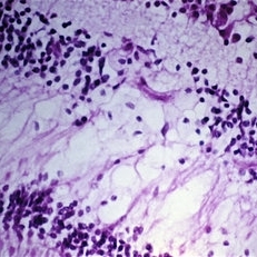
Deep retinal exudate. There is accumulation of lipid-laden histiocytes in the outer plexiform and inner nuclear layers of the retina in the posterior pole area in diabetic retinopathy.
Condition/keywords: diabetic retinopathy, histiocytes, retinal exudates
-
 Slide 9-18
Feb 26 2019 by Lancaster Course in Ophthalmology
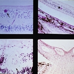
Malignant hypertension with retinal arterioles that are thickened and have fibrinoid necrosis (arrows). Retinal exudates (asterisk) and papilledema are also present. Papilledema is evidenced by fullness of the optic nerve head and peripapillary crowding of the retina (lower right).
Condition/keywords: fibrinoid, malignant hypertension, papilledema, retinal arteriole, retinal exudates
-
 Slide 9-19
Feb 26 2019 by Lancaster Course in Ophthalmology
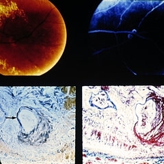
Retinal arterial macroaneurysm. A ring of retinal exudate partially surrounds the macroaneurysm (upper left), which is more clearly delineated by fluorescein (upper right). The retinal arteriole is greatly dilated, and the stain for elastic tissue shows a localized area of disruption and loss of the internal elastic membrane (arrow). The surrounding retina is thickened by edema and some hemorrhage. The ectatic area of the vessel wall is greatly thickened by the accumulation of a laminated fibrinous material. (Courtesy of Alan Friedman, M.D.)
Condition/keywords: retinal arterial macroaneurysm, retinal exudates
-
---thumb.jpg/image-square;max$300,300.ImageHandler) Yellow White Subretinal Exudates
Yellow White Subretinal Exudates
Feb 13 2013 by From the Collections of Thomas M. Aaberg, MD and Thomas M. Aaberg Jr., MD
Yellow white subretinal exudates/lesions in macula and mid periphery.
Condition/keywords: macula, subretinal exudates
 Loading…