Search results (43 results)
-
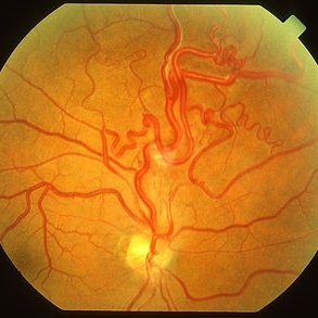
 Retinal Arteriovenous Malformations (Racemose Hemangiomatosis)
Mar 30 2018 by Rameez N Hussain, MD
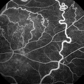
A 7-years-old Portuguese girl with unilateral retinal arteriovenous malformations composed of dilated, tortuous vessels with normal vision.
Photographer: Thambi Durai. Consultant Optometrist, Orbit Health Care - Dr Agarwal's Eye Hospital, Maputo, Mozambique
Imaging device: TOPCON
Condition/keywords: racemose hemangioma, retinal arteriovenous malformations, Wyburn-Mason
-
 Wyburn-Mason Syndrome (Racemose Hemangiomatosis)
Mar 30 2018 by Rameez N Hussain, MD
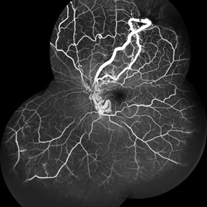
A 7-year-old Portuguese girl with unilateral retinal arteriovenous malformations composed of dilated, tortuous vessels with normal vision.
Photographer: Thambi Durai
Imaging device: TOPCON
Condition/keywords: arteriovenous malformation, racemose hemangioma, Wyburn-Mason
-
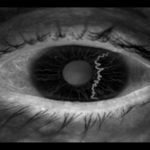 Iris Racemose Hemangioma
Jan 1 2023 by Maxwell J Wingelaar, MD
Fluorescein Angiogram of 66 year old female presented with an iris racemose hemangioma
Photographer: Ken Huff
Condition/keywords: Racemose hemangioma
-
 Wyburn Mason Syndrome
May 2 2013 by Henry J. Kaplan, MD
Racemose angioma of the retina in Wyburn Mason syndrome.
Condition/keywords: racemose hemangioma
-
 Wyburn-Mason Syndrome (Racemose Angioma)
Mar 23 2024 by Pushkar Mahale
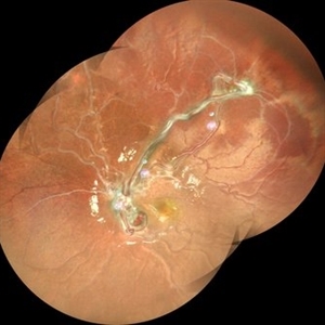
Fundus photograph of a 10 year old child presenting with no perception of light in right eye. Fundus examination revealed dilated and tortuous retinal vessels suggestive of Racemose Hemangioma.
Photographer: Dr Pushkar Mahale
Condition/keywords: racemose hemangioma, Wyburn -Mason Syndrome
-
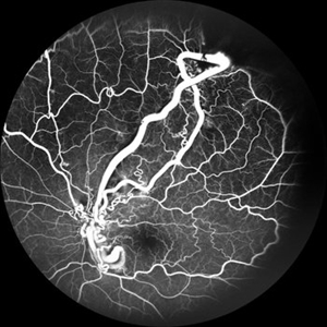
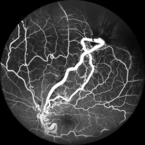 From Artery to Vein, No Detour: Meet the AV Maverick: Racemose Hemangioma
Jul 1 2025 by rohan jain
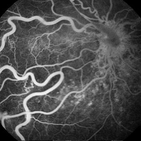
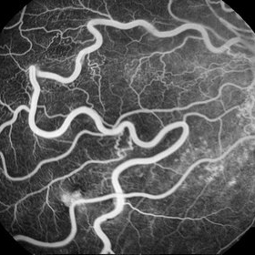
A case of 10 year old girl with defective vision in LE (6/60) who presented us with this condition.
Photographer: Dr. ROHAN JAIN
Imaging device: mirante
Condition/keywords: arteriovenous malformation, FFA in a case of Racemose angioma, racemose hemangioma
-
 Peri-papillary Vascular Loop
Jun 2 2020 by Dhaivat Shah
Peri-papillary vascular loops (PVL) are rare congenital vascular malformations, which are usually detected as accidental finding during routine fundus examination. They can often be confused with tributary vein occlusion or racemose hemangioma. Although benign and asymptomatic, they can be rarely associated with vitreous hemorrhage and arterial occlusion. We herein present a case of a 60-year-old hypertensive male, who was diagnosed elsewhere to have a tributary vein occlusion and was referred to us. FFA was advised to rule out neovascularization, surrounding capillary non perfusion and mass lesion (hemangioma). On FFA, the arterial loop showed a slightly delayed filling (3-5 seconds) as compared to the other arterial vessels and the original vessel appeared to be a branch arising from central retinal artery. The choroidal filling was delayed in the area supplied by the loop. A cilioretinal artery was also noted. The patient was diagnosed to have a Peri-papillary vascular arterial loop (PVL), likely to be congenital in origin. The patient was reassured and was advised yearly follow up. These loops are usually accidental findings discovered during routine fundus examination. Since these vessels are looped and tortuous, they exhibit a slower and laminar blood flow, which make them more prone for arterial occlusions. The vitreous in this area tends to be adherently attached, so during PVD induction, it is likely to cause a tear and hemorrhage leading to vitreous hemorrhage. Until and unless there is a break, this hemorrhage tends to resolve on its own and does not warrant treatment. If there is an evident break, it can be dealt with laser barrage.
Photographer: Choithram Netralaya
Condition/keywords: congenital prepapillary vascular loop
-
 Racemose Hemangioma
Feb 20 2013 by From the Collections of Thomas M. Aaberg, MD and Thomas M. Aaberg Jr., MD
Dilated tortuous blood vessels.
Condition/keywords: racemose hemangioma
-
---thumb.jpg/image-square;max$300,300.ImageHandler) Racemose Hemangioma
Racemose Hemangioma
Feb 20 2013 by From the Collections of Thomas M. Aaberg, MD and Thomas M. Aaberg Jr., MD
dilated vessels
Condition/keywords: racemose hemangioma
-
 Racemose Hemangioma
Mar 14 2021 by Luiz A Zago, PhD
Racemose hemangioma in a 30-year-old woman with ambliopia in this eye.
Photographer: Luiz Alberto Zago Filho
Imaging device: Topcon 50IX
Condition/keywords: racemose hemangioma
-
 Racemose Hemangioma and Retinal Vein Occlusion
Mar 5 2013 by Eduardo Torres-Porras, MD
A 13-year-old woman had a history of decreased vision for 2 years. Visual acuity was 20/400.
Photographer: Camelia Rosales Lara
Condition/keywords: retinal angiomatous proliferation (RAP)
-
 Racemose Hemangioma and Retinal Vein Occlusion
Mar 5 2013 by Eduardo Torres-Porras, MD
A 13-year-old woman had a history of decreased vision for 2 years. Visual acuity was 20/400.
Photographer: Camelia Rosales Lara
Condition/keywords: retinal angiomatous proliferation (RAP)
-
 Racemose Hemangiomatosis
May 27 2020 by Jamin S. Brown, MD
Fundus photo of 25-year-old female with racemose hemangiomatosis OS.
Photographer: Stefanie Palmer CRA, Retina-Vitreous Surgeons of CNY
Condition/keywords: racemose hemangioma
-
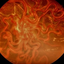
 Wyburn-Mason (Raecemose Hemangioma)
Oct 2 2013 by Jerald A. Bovino, MD
The patieint has typical Wyburn Mason syndrome with dilated, extrement tortuous venules.
Condition/keywords: dilated and tortous veins, racemose hemangioma
-
 Wyburn-Mason Syndrome
Jan 28 2013 by Ingrid E. Zimmer-Galler, MD
Fundus photographs and fluorescein angiogram of a 29-year-old male with asymptomatic racemose lesions characterized by direct artery-to-vein communication.
Photographer: David Emmert, Wilmer Eye Institute, Johns Hopkins University
Condition/keywords: racemose hemangioma, retinal arteriovenous malformations
-
 Retinal Arteriovenous Malformations (Racemose Hemangiomatosis)
Mar 30 2018 by Rameez N Hussain, MD
A 7-year-old Portuguese girl with unilateral retinal arteriovenous malformations composed of dilated, tortuous vessels with normal vision.
Photographer: Thambi Durai, Consultant Optometrist, Orbit Health Care - Dr Agarwal's Eye Hospital, Maputo, Mozambique
Imaging device: TOPCON
Condition/keywords: racemose hemangioma, retinal arteriovenous malformations, Wyburn-Mason
-
 Wyburn-Mason Syndrome
Feb 20 2015 by H. Michael Lambert, MD
Racemose hemangiomatosis; Large arteriovenous malformation in the right eye.
Condition/keywords: arteriovenous malformation, racemose hemangioma, Wyburn-Mason
-
 Racemose Hemangioma and Retinal Vein Occlusion
Mar 5 2013 by Eduardo Torres-Porras, MD
A 13-year-old woman had a history of decreased vision for 2 years. Visual acuity was 20/400.
Photographer: Camelia Rosales Lara
Condition/keywords: retinal angiomatous proliferation (RAP)
-
 From Artery to Vein, No Detour: Meet the AV Maverick: Racemose Hemangioma
Jul 1 2025 by rohan jain
A case of 10 year old girl with defective vision in LE (6/60) who presented us with this condition.
Photographer: Dr. ROHAN JAIN
Imaging device: mirante
Condition/keywords: arteriovenous malformation, FFA in a case of Racemose angioma, racemose hemangioma
-
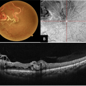 Multimodal Imaging of a Type 3 Retinal Racemose Hemangioma
Sep 8 2024 by Maria Antonia Orrego
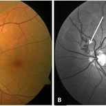
We present the case of a 33 year-old woman with visual loss of her left eye since childhood. Fundus examination revealed a retinal arteriovenous malformation with vessels originating from the optic nerve and extending to the fovea and equator, corresponding to a type 3 retinal racemose hemangioma (A). Infrared reflectance imaging confirmed findings described in funduscopy (B). Spectral domain optical coherence tomography shows dilated vessels in the internal and external retinal layers and adjacent intraretinal fluid (C).
Photographer: Dr. Maria Antonia Orrego V, Universidad CES, Clinica Clofán, Medellín, Colombia
Imaging device: Optovue Solix
Condition/keywords: arteriovenous malformation, multimodal imaging, racemose hemangioma, retinal arteriovenous malformations
-
 Racemose Hemangioma
Feb 20 2013 by From the Collections of Thomas M. Aaberg, MD and Thomas M. Aaberg Jr., MD
Dilated tortuous blood vessels.
Condition/keywords: racemose hemangioma
-
 Racemose Hemangioma
Apr 7 2022 by Sengul Ozdek, MD, FEBO, FASRS
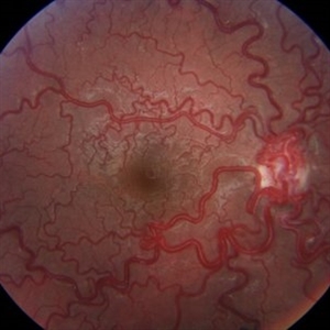
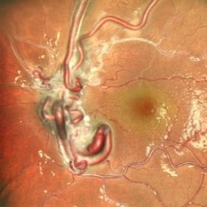
This is a fundus picture of a case with Racemose hemangioma. It is a rare sporadic congenital arteriovenous malformation, with unilateral involvement. It is characterized by a dilated, tortuous, tangled network of retinal vessels, mostly emerging from the optic disc and extending towards periphery, with no distinction between arterioles and venules.
Photographer: Refiye Basdogan
Imaging device: Canon
Condition/keywords: congenital arteriovenous malformation, Racemose hemangioma
 Loading…