Search results (18 results)
-
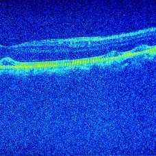
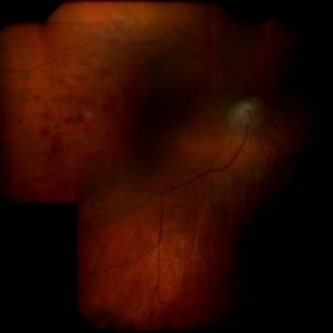
 Retained Perfluorcarbon and Macular Edema After Silicon Oil Removal 3D
Jul 24 2017 by Nelson Chamma Capelanes, MD
SD-OCT and HRA from a 42-year-old patient after silicon oil removal. The image shows macular edema and retained perfluorcarbon.
Photographer: Nelson Chamma Capelanes, Promedica Indaiatuba, Brazil
Condition/keywords: macular edema, post-vitrectomy, retained perfluorocarbon
-
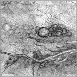
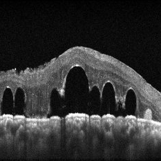 submacular perfluorocarbon liquid
Sep 7 2022 by JEFFERSON R SOUSA, Tecg.º (Biomedical Systems Technology)
A 63-year-old male patient underwent vitreoretinal surgery with the use of perfluorocarbon. From a technological point of view, extended-field retinography presents many points of focus variation due to the difficulty of establishing a diffuse focus, as it is a recent post-operative case. In OCT Fundus Enface, although it has a low resolution, it is extremely important for documenting the presence of perfluor. Best seen in structural OCT.
Photographer: JEFFERSON ROCHA DE SOUSA - Retinal Department at Instituto Dr. Suel Abujamra Sao Paulo-Brazil
Imaging device: Optical Coherence Tomography system OCT CIRRUS 5000, Protocol, HD 5 Line
Condition/keywords: perfluorocarbon fluid, post-vitrectomy, submacular perfluorocarbon liquid (PFO), vitrectomy
-
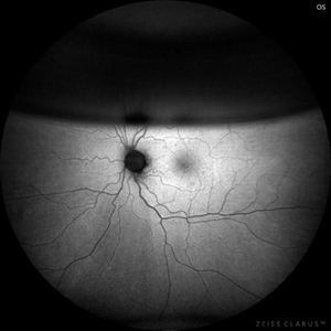
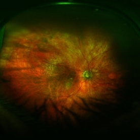 Optic Atrophy and Attenuated Retinal Vessels Following Endophthalmitis
Jul 12 2014 by Philip J. Polkinghorne, MD
This elderly lady underwent a vitrectomy for post-surgical endophthalmitis. The infection was successfully treated but the functional outcome was poor because of optic atrophy and attenuated retinal vessels.
Photographer: Alex Fraser
Imaging device: Optos Camera
Condition/keywords: attenuated vessels, endophthalmitis, optic atrophy, post-vitrectomy
-
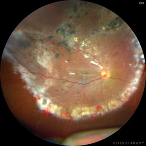 submacular perfluorocarbon liquid
Sep 7 2022 by JEFFERSON R SOUSA, Tecg.º (Biomedical Systems Technology)
A 63-year-old male patient underwent vitreoretinal surgery with the use of perfluorocarbon. From a technological point of view, extended-field retinography presents many points of focus variation due to the difficulty of establishing a diffuse focus, as it is a recent post-operative case. In OCT Fundus Enface, although it has a low resolution, it is extremely important for documenting the presence of perfluor. Best seen in structural OCT.
Photographer: JEFFERSON ROCHA DE SOUSA - Retinal Department at Instituto Dr. Suel Abujamra Sao Paulo-Brazil
Imaging device: Clarus 700 - Zeiss, 135 degree images.
Condition/keywords: perfluorocarbon fluid, post-vitrectomy, submacular perfluorocarbon liquid (PFO), vitrectomy
-
 Intravitreal Triamcinolone and Gas
Feb 25 2017 by William G. Campbell, MD
Post-operative photograph of left fundus 10 days following vitrectomy with triamcinolone injection and SF6 gas tamponade.
Photographer: Tanya Pejnovic, Melbourne Retina Associates, Melbourne, Australis
Imaging device: Optos 200 Tx wide-field camera
Condition/keywords: post-vitrectomy
-
 9 in a Row
Oct 30 2023 by SHAILEEN PARIKH, MS, DO, FMRF
Fundus Photograph of RE showing multiple Pigment Epithelial Detachments in a 1 month post operative patient who underwent surgery for Temporal retinal detachment with attached macula
Photographer: Dr. Aneri Ambani
Condition/keywords: pigment epithelial detachment (PED), post-vitrectomy
-
 Giant Retinal Tear Post-Vitrectomy
May 8 2021 by ? ?
Fundus photograph of a 30-year-old woman post-vitrectomy and laser.
Condition/keywords: giant retinal tear, post CE, post-vitrectomy
-
 Intraocular Gas bubble - Wide field Fundus Autofluorescence
Sep 6 2021 by Ricardo Leitão Guerra
Wide field confocal scanning laser ophthalmoscopy 7 days after vitrectomy to treat a macular hole.
Imaging device: Zeiss Clarus 700
Condition/keywords: gas bubble, macular hole, pars plana vitrectomy (PPV), post-vitrectomy, vitrectomy
-
 Intraocular Gas Bubble - Wide-field True Color CSLO
Sep 6 2021 by Ricardo Leitão Guerra
Wide field confocal scanning laser ophthalmoscopy 7 days after vitrectomy to treat a macular hole.
Imaging device: Zeiss Clarus 700
Condition/keywords: gas bubble, macular hole, pars plana vitrectomy (PPV), post-vitrectomy, vitrectomy
-
 Multiple Pigment Epithelial Detachments Post Vitrectomy Surgery
Oct 30 2023 by SHAILEEN PARIKH, MS, DO, FMRF
OCT of RE showing '4 in a Row' Pigment Epithelial Detachments in a 1 month post operative patient who underwent surgery for Temporal retinal detachment with attached macula
Photographer: Dr. Aneri Ambani
Condition/keywords: pigment epithelial detachment (PED), post-vitrectomy
-
 Post Vitrectomy Color Fundus Photo Showing Resolution of Hemorrhage
Aug 10 2014 by Thomas A. Ciulla, MD, MBA, FASRS
She underwent pars plana vitrectomy, submacular tissue plasminogen activator injection, gas injection, face down positioning. Follow up 3 months later showed VA: 20/80 OS. Color fundus photos show resolution of hemorrhage.
Condition/keywords: post-vitrectomy, submacular hemorrhage, tissue plasminogen activator (tPA), wet age-related macular degeneration (wet AMD)
-
 Post Vitrectomy Early Phase Angiogram
Aug 10 2014 by Thomas A. Ciulla, MD, MBA, FASRS
She underwent pars plana vitrectomy, submacular tissue plasminogen activator injection, gas injection, face down positioning. Follow up 3 months later showed VA: 20/80 OS. Angiography shows resolution of submacular hemorrhage, no active CNVM, and no retinal artery macroaneurysm.
Condition/keywords: post-vitrectomy, submacular hemorrhage, tissue plasminogen activator (tPA), wet age-related macular degeneration (wet AMD)
-
 Post Vitrectomy Late Phase Angiogram
Aug 10 2014 by Thomas A. Ciulla, MD, MBA, FASRS
She underwent pars plana vitrectomy, submacular tissue plasminogen activator injection, gas injection, face down positioning. Follow up 3 months later showed VA: 20/80 OS. Angiography shows resolution of submacular hemorrhage, no active CNVM, and no retinal artery macroaneurysm.
Condition/keywords: post-vitrectomy, submacular hemorrhage, tissue plasminogen activator (tPA), wet age-related macular degeneration (wet AMD)
-
 Post Vitrectomy OCT Showed Atrophy and No Exudation
Aug 10 2014 by Thomas A. Ciulla, MD, MBA, FASRS
Post vitrectomy OCT showed atrophy and no exudation. VA 20/80.
Condition/keywords: post-vitrectomy, submacular hemorrhage, tissue plasminogen activator (tPA), wet age-related macular degeneration (wet AMD)
-
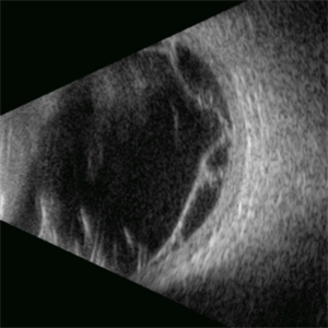 Proliferative Vitreoretinopathy
Apr 17 2025 by Gustavo Uriel Fonseca Aguirre
This B-mode transverse ultrasound scan depicts a post-vitrectomy eye with recurrent retinal detachment in a patient with diabetic retinopathy history. The image reveals fresh vitreous cavity hemorrhage and subretinal bleeding, along with subretinal proliferative bands (PVR strands). These findings indicate complicated tractional re-detachment with active hemorrhagic components.
Photographer: Gustavo U. Fonseca Aguirre, Hospital Conde de Valenciana, Ciudad de México
Condition/keywords: proliferative vitreoretinopathy (PVR)
-
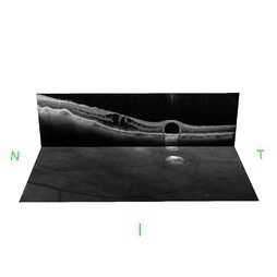
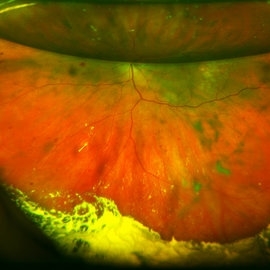
 Retained Perfluorcarbon and Macular Edema After Silicon Oil Removal
Jul 24 2017 by Nelson Chamma Capelanes, MD
SD-OCT and HRA from a 42-year-old patient after silicon oil removal. The image shows macular edema and retained perfluorcarbon.
Photographer: Nelson Chamma Capelanes, Promedica Indaiatuba, Brazil
Condition/keywords: macular edema, post-vitrectomy, retained perfluorocarbon
-
 submacular perfluorocarbon liquid
Sep 7 2022 by JEFFERSON R SOUSA, Tecg.º (Biomedical Systems Technology)
A 63-year-old male patient underwent vitreoretinal surgery with the use of perfluorocarbon. From a technological point of view, extended-field retinography presents many points of focus variation due to the difficulty of establishing a diffuse focus, as it is a recent post-operative case. In OCT Fundus Enface, although it has a low resolution, it is extremely important for documenting the presence of perfluor. Best seen in structural OCT.
Photographer: JEFFERSON ROCHA DE SOUSA - Retinal Department at Instituto Dr. Suel Abujamra Sao Paulo-Brazil
Imaging device: Optical Coherence Tomography system OCT CIRRUS 5000, Protocol OCT Fundus Enface.
Condition/keywords: perfluorocarbon fluid, post-vitrectomy, submacular perfluorocarbon liquid (PFO), vitrectomy
-
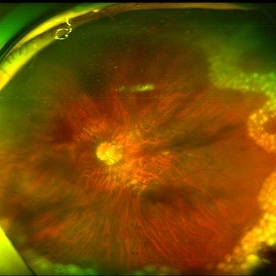
 Vasculitis Following Successful Treatment of Endophthalmitis
Jul 12 2014 by Philip J. Polkinghorne, MD
A 65-year-old lady presented with endophthalmitis following cataract surgery. She had a vitrectomy and intravitreal antibiotics. Post-operatively she did well. The montage shows persisting vasculitis and retinal haemorrhages three months following presentation.
Condition/keywords: endophthalmitis, post-vitrectomy
 Loading…