Search results (304 results)
-
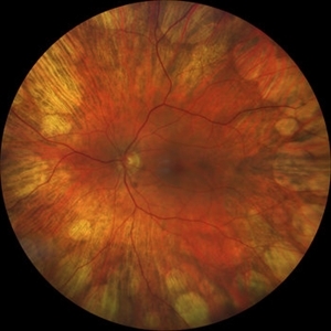
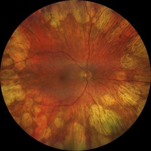
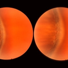
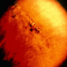
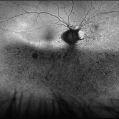 Phoenix
Feb 21 2024 by Sayena . Jabbehdari, MD, MPH, MBA
A 60-year-old Caucasian female presented with reduced night vision and constricted visual fields. The fundus exam revealed pigmentary changes in the peripheral retina. Fundus autofluorescence depicted the schematic appearance of a Phoenix , with the hypo-autofluorescence corresponding to the head and wings of the phoenix. Genetic testing was positive for a heterozygous RHO mutation
Photographer: Sayena Jabbehdari MD MPH
Condition/keywords: retinitis pigmentosa
-
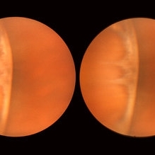
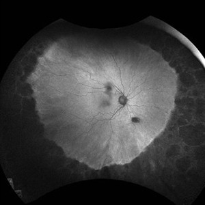
 The Peripheral Retina in Profile: A Stereoscopic Atlas
Mar 12 2013 by Norman Byer
The stereoscopic atlas contains unique stereo photographs vividly portraying the changes in the peripheral fundus and their histopathology, incidence and risks.
Condition/keywords: stereo pair, video
-
 Didanosine Toxicity
Jan 27 2020 by Nimesh A. Patel, MD, FASRS
Patient with history of HIV treated with didanosine. Developed gyrate like peripheral retinal atrophy with central sparing. Vision is 20/25
Imaging device: Clarus
Condition/keywords: AIDS, didanosine, HIV
-
 Didanosine Toxicity
Jan 27 2020 by Nimesh A. Patel, MD, FASRS
Patient with history of HIV treated with didanosine. Developed gyrate like peripheral retinal atrophy with central sparing. Vision is 20/25
Imaging device: Clarus
Condition/keywords: AIDS, didanosine, HIV
-
 Normal Nasal Ora Serrata
Nov 9 2012 by Norman Byer
This shows the normal nasal ora serrata. Note the dentate processes which divide the nasal ora into prominent bays and teeth
Condition/keywords: dentate processes, normal nasal ora serrata, ora bay, ora teeth
-
 Normal Temporal Ora Serrata
Nov 9 2012 by Norman Byer
This is the normal temporal ora serrata in a 26-year-old man. Note the typical ragged moth-eaten appearance caused by peripheral cystoid degeneration. This appearance may be present in infants but is always present beyond the age of eight years.
Condition/keywords: ora serrata, peripheral cystoid degeneration
-
 Snail Track Peripheral Retinal Degeneration
Apr 29 2022 by Otakar Dušek, M.D. Ph.D.
Colour fundus photograph of 22-year-old woman with incidentally found snail track retinal degeneration in the superior temporal periphery of the retina of the right eye.
Photographer: Otakar Dušek, Charles University, Prague
Imaging device: Zeiss Clarus
Condition/keywords: peripheral retinal degeneration
-
 Sudden Posterior Vitreous Detachment
Nov 9 2012 by Norman Byer
This 60-year-old man suffered a sudden posterior vitreous detachment which produced a large tractional retinal tear at 11:30 o’clock in this eye. This white cystic retinal tuft located at 9:30 also suffered minor injury at the same time as revealed in the next slide pair.
Condition/keywords: posterior vitreous detachment, white retinal tuft
-
 Alagille Syndrome UWF Autofluorescence
Dec 4 2023 by Isaac Ezon, MD
43 yo Female with known Alagille Syndrome, referred for peripheral retinal changes. Subjective nyctalopia but no other symtpoms. Alagille Syndrome UWF Autofluorescence.
Photographer: Tara Murray
Imaging device: Optos
Condition/keywords: hereditary choroidal dystrophy, hereditary retinal degeneration
-
 Commotio Retinae
Aug 7 2025 by Gabriel Costa Andrade, PhD
Color fundus photograph of a 13-year-old girl who was hit by accidental discharge of gel bullet in her right eye. She presented with retinal whitening with intraretinal hemorrhages in temporal inferior area of the peripheral retina.
Photographer: Gabriel Andrade
Condition/keywords: macula, Retina, Trauma
-
 Congenital Meridional
Nov 9 2012 by Norman Byer
This is the same case as seen in the previous photograph but showing an area just below the lower end of the dialysis. It shows a congenital meridional fold at the 2 o’clock meridian with a retinal break at the posterior end possibly caused by the direct injury described previously.
Condition/keywords: meridional fold, ora serrata, retinal break
-
 Elevated Lesion
Nov 9 2012 by Norman Byer
This photograph and the next are two views of a very interesting elevated lesion in a 45-year-old man. This photograph shows the immense value of closely scrutinizing the profile of the indented area. Note that in the middle of the slide there is a sudden break in the continuity of the dark convex shadow that lies just behind the crest of the scleral indentation. If the elevated tissue is "filmy" or "wispy" or filamentous as in this case, it raises a strong suspicion that a retinal break is present just behind it.
Condition/keywords: elevated retinal lesion, elevated tissue, retinal break, scleral indentation
-
 Gyrate Atrophy
Oct 15 2022 by Maxwell J Wingelaar, MD
Fundus photograph of a 15-year-old male with peripheral retinal changes consistent with gyrate atrophy
Photographer: Jarrod Wehmeier
Condition/keywords: gyrate atrophy
-
 Laser Photocoagulation
Nov 9 2012 by Norman Byer
This shows the same lesion four days after laser photo coagulation. The new hemorrhages seen in this photograph did not occur during the photocoagulation but developed within the next four days.
Condition/keywords: argon photocoagulation, laser photocoagulation, preretinal hemorrhage
-
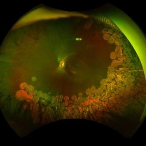
 Lattice Degeneration
May 2 2013 by Henry J. Kaplan, MD
Pigmented lattice degeneration with lattice "wicker" caused by sclerotic blood vessels.
Condition/keywords: lattice degeneration, peripheral retinal degeneration
-
 Lattice Lesion
Nov 9 2012 by Norman Byer
This lattice lesion in a 36-year-old woman has remained unchanged over a period of 13 years. It shows a moderate snailtrack feature with discrete yellow dots visible on the surface of the lesion and especially along the posterior border. One of these can be well seen just below the lesion superimposed over the dark shadow of the scleral indentation. The exact nature of these yellow dots is still not entirely clear.
Condition/keywords: lattice degeneration, moderate snail track, scleral indentation, yellow dots
-
 Normal Nasal Ora Serrata
Nov 9 2012 by Norman Byer
This is the normal nasal ora serrata showing a prominent meridional fold. Such folds are most commonly seen at the lower part of the upper nasal quadrant, and are present in 26% of the population. They are a normal developmental variation and are often bilateral.
Condition/keywords: meridional fold, normal developmental variation, normal nasal ora serrata, upper nasal quadrant
-
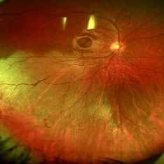
 Peripheral Retinal Degeneration (L-ORD)
Apr 17 2024 by Virginia Gebhart
92 year old female with bilateral patchy, sharply demarcated circular areas of chorioretinal atrophy with hyperpigmented margins in the mid to far periphery. Labs showed normal plasma ornithine levels ruling out generalized gyrate atrophy. Also intermediate uveitis and CMD/CME. FTA-ABS, Quant gold, and HLA-A29 labs all negative.
Photographer: Virginia Gebhart
Imaging device: Optos California
Condition/keywords: cystoid macular degeneration, cystoid macular edema (CME), FA, Fluorescein angiography, peripheral retinal degeneration
-
Peripheral Retinal Hole with OCT Co-localization
Sep 26 2023 by Bradley T. Smith, MD, FASRS
Peripheral asymptomatic atrophic retinal hole with OCT co localization demonstrating small cuff of sub retinal fluid. Near infrared imaging shows hyper reflectivity through hole.
Condition/keywords: atrophic hole, lattice degeneration, OCT
-
 Proliferative Diabetic Retinopathy
Oct 15 2012 by Susanna S. Park, MD, PhD
Fluorescein angiogram of the left eye of a 65 year old woman with diabetes mellitus showing nasal peripheral retinal capillary dropout and neovascularization of the disc. Scattered retinal microaneurysms are also noted
Photographer: Ellen Redenbo, University of California Davis Eye Center
Imaging device: Optos
Condition/keywords: proliferative diabetic retinopathy (PDR)
-
 Retinal Break
Nov 9 2012 by Norman Byer
This is the right eye of a 49-year-old woman showing a tiny retinal break adjacent to the temporal ora serrata. It has remained exactly the same without treatment for nine years.
Condition/keywords: ora serrata, retinal break
-
 Retinal Detachment
Nov 9 2012 by Norman Byer
This 18-year-old girl gave the history of having been hit in this eye three years before with a fist and of having retinal surgery nine months previously, which was temporarily successful. When the photograph was taken, she had a total left retinal detachment with a small nasal dialysis which had not been treated. She also had two prominent intraretinal cysts, one of which is shown here. The retina promptly reattached following further surgery and the next slide shows an interesting change in this cyst.
Condition/keywords: intraretinal cyst, small nasal dialysis
-
 Retinal Lesion
Nov 9 2012 by Norman Byer
This 30-year-old man sustained a severe blow to his brow region which resulted in a variety of injuries including hyphema and vitreous and retinal hemorrhages. This photograph shows a retinal lesion, which is either a tiny area of elevated full thickness retina or a traumatic retinal cyst.
Condition/keywords: elevated retinal lesion, retinal hemorrhage, traumatic retinal cyst
-
 Retinalschisis
Nov 9 2012 by Norman Byer
This 43-year-old woman had the small area of retinalschisis and associated small localized retinal detachment resulting from breaks in both retinal layers when she was first examined. This eye was asymptomatic and has remained exactly the same during two years of observation without treatment. In this picture, the outer layer break is demarcated by the yellow line. The inner layer is seen intact over this hole but has itself two small holes in the upper part of the photograph. Thus, it is evident that even retinoschisis with double layer holes does not necessarily progress to a clinical retinal detachment. The viscous nature of the fluid in the retinoschisis cavity is probably a contributing factor in this non progressive tendency.
Condition/keywords: inner layer holes, outer layer hole, retinoschisis
-
 Rhegmatogenous retinal detachment with dislocated IOL in a Morning Glory anomaly
Jul 27 2023 by Gustavo Aguirre-Suarez
Fundus photograph of a 13-year-old male with a history of congenital cataract surgery in his right eye in 2019. The patient presents with sudden visual loss. Upon examination, a dislocated IOL is observed in the posterior segment, accompanied by a rhegmatogenous retinal detachment featuring peripheral retinal tears and horseshoe breaks. Additionally, a morning glory disc anomaly is also present in this patient.
Photographer: Gustavo Aguirre-Suarez
Imaging device: Mirante, NIDEK
Condition/keywords: dislocated posterior chamber intraocular lens (PCIOL), Morning Glory Anomaly, rhegmatogenous retinal detachment
 Loading…