Search results (80 results)
-
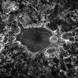
 PRP laser
Mar 29 2013 by Henry J. Kaplan, MD
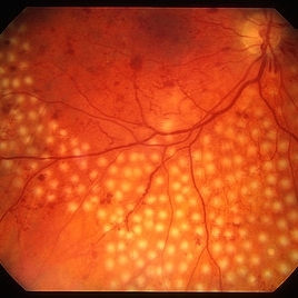
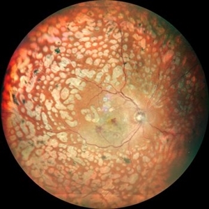
Right after PRP laser in PDR.
Condition/keywords: laser photocoagulation, pan-retinal photocoagulation (PRP)
-
 Sickle Cell Retinopathy
Feb 15 2021 by Kim Barrett
24-year-old female with Sickle Cell Retinopathy, stage 3. She confirms she has the trait as well as her grandmother, mother and a sibling. She has seafan neovascularization superotemporal OD. Current VA is 20/20. Photo is pre-PRP laser with areas of non-profusion temporally.
Photographer: Kim Barrett C.O.A. Retina Specialist of Michigan, Grand Rapids, MI
Imaging device: Optos California
Condition/keywords: neovascularization (NV), pan-retinal photocoagulation (PRP), sickle cell retinopathy, stage 3, trait
-
 Active diabetic retinopathy despite PRP
Oct 30 2022 by Diego Andrés Rodriguez, MD
A 52-year-old patient with active proliferative diabetic retinopathy despite good glycemic control and PRP performed 1 year ago in the right eye
Photographer: Sociedad de Cirugía Ocular
Imaging device: Clarus 700
Condition/keywords: diabetic retinopathy, pan-retinal photocoagulation (PRP), proliferative diabetic retinopathy (PDR), wide angle imaging
-
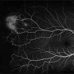
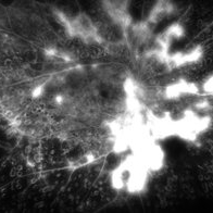
 Central Retinal Vein Occlusion with Retinal Neovascularization
Jan 19 2022 by Olivia Rainey
Ultra-widefield fluorescein angiogram of a 56-year-old male with a Central Retinal Vein Occlusion with Retinal Neovascularization affecting his left eye. The patient presented on 1/19/2022 with scNLP vision in the left eye. The patient has good PRP, however areas of ischemia still remain untreated by laser. He also has severe neovascular glaucoma contributing to his poor vision.
Photographer: Olivia Rainey, OCT-C, COA
Imaging device: Optos California
Condition/keywords: central retinal vein occlusion (CRVO), FA early phase, hemorrhage, ischemic CRVO, left eye, neovascular glaucoma, Optos, pan-retinal photocoagulation (PRP), retinal ischemia, retinal neovascularization, ultra-wide field imaging
-
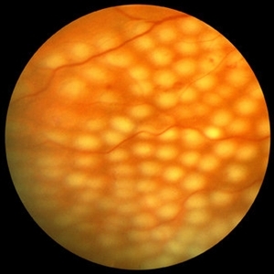
 CNVM in Pan-retinal Photocoagulated Patient
Dec 30 2020 by ASRS Staff
Wide fundus photograph of 65-year-old, female, diabetic patient.
Imaging device: Nidek Mirante
Condition/keywords: age-related macular degeneration (AMD), diabetes, pan-retinal photocoagulation (PRP)
-
 Coats' Disease
Jul 10 2018 by Karen Panzegrau
Ultra-wide field images of a 30-year-old male with Coats' Disease affecting his right eye. Patient had sectoral PRP with significant improvement in lipid after 8 months of being lost to folllow up. Vision has improved beyond expectations given severity of lipid.
Photographer: Karen Panzegrau
Imaging device: Optos
Condition/keywords: Coats' disease, fundus photograph, lipid exudation, pan-retinal photocoagulation (PRP)
-
 Diabetic Retinopathy
Oct 18 2012 by Raj K. Maturi, MD
Photographer: Tom Steele, CRA
Imaging device: Optos
Condition/keywords: pan-retinal photocoagulation (PRP)
-
 Diabetic Retinopathy
Oct 18 2012 by Raj K. Maturi, MD
Photographer: Tom Steele, CRA
Imaging device: Optos
Condition/keywords: pan-retinal photocoagulation (PRP)
-
 Diabetic Retinopathy Treated with PRP Laser
Jun 8 2021 by Ronald Coriasso
Diabetic retina treated with complete 360 PRP laser, taken during fluorescein angiogram.
Photographer: Ronald Coriasso
Imaging device: OPTOS
Condition/keywords: pan-retinal photocoagulation (PRP)
-
 Diabetic Tractional Retinal Detachment
Jan 23 2019 by Olivia Rainey
Ultra-wide field pseudocolor image of an 43-year-old female with a diabetic tractional retinal detachment and a vitreous hemorrhage affecting her right eye.
Photographer: Olivia Rainey
Imaging device: Optos
Condition/keywords: diabetes, diabetic traction detachment, Optos, pan-retinal photocoagulation (PRP), proliferative diabetic retinopathy (PDR), pseudocolor, ultra-wide field imaging, vitreous hemorrhage
-
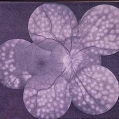
 Extensive Pan-Retinal Photocoagulation
Apr 19 2013 by Brandon G. Busbee, MD
Extensive pan-retinal photocoagulation.
Photographer: Alecia Camp, CRA - Tennessee Retina - Nashville, TN
Imaging device: Topcon TRC 50-EX
Condition/keywords: neovascularization (NV), pan-retinal photocoagulation (PRP)
-
 Exudative Macular Detachment After Intensive Laser Photocoagulation
Mar 12 2016 by Sjakon G Tahija, MD
Fundus photograph of 44-year-old man with exudative detachment of the macula after vitrectomy and ILM peeling for proliferative diabetic retinopathy combined with intensive endolaser photocagulation.
Photographer: Avris Siahaan, Klinik Mata Nusantara
Condition/keywords: exudative detachment, pan-retinal photocoagulation (PRP)
-
 Neovascularization of the Disc
Apr 19 2013 by Brandon G. Busbee, MD
Persistant NVD after extensive PRP.
Photographer: Alecia Camp, CRA - Tennessee Retina - Nashville, TN
Imaging device: Topcon TRC 50-EX
Condition/keywords: neovascularization of the disc (NVD), pan-retinal photocoagulation (PRP)
-
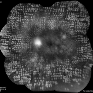
 Pan-Retinal Photocoagulation
Apr 5 2018 by Mohamed Tawfik, MD
Wide field FFA post phaco vitrectomy of a case of vitreous hemorrhage show PRP with regression of diabetic retinopathy.
Photographer: Mohamed A,Tawfik MD,FRCSed
Condition/keywords: pan-retinal photocoagulation (PRP)
-
 Serous Retinal Detachment in Advanced Proliferative Diabetic Retinopathy
Feb 15 2024 by Annaka Gooding
Ultra-Wide fundus photograph of a 29 year old female with a Serous Retinal Detachment in Advanced PDR. Patient present to clinic with LP vision following PPV and fill in PRP. Physician recommended oral prednisone treatment and to reassess at their following visit.
Photographer: Annaka Gooding, CPO
Imaging device: Optos California RGB
Condition/keywords: Diabetes, diabetic macular edema, fundus photography, OPTOS CALIFORNIA, pan-retinal photocoagulation (PRP), pars plana vitrectomy (PPV), proliferative diabetic retinopathy (PDR), serous retinal detachment, ultra-wide field imaging
-
 PDR; High Myopia; PRP
May 2 2019 by Carissa Hurdstrom
PDR; high myopia; PRP
Imaging device: Optos
Condition/keywords: high myopia, pan-retinal photocoagulation (PRP), proliferative diabetic retinopathy (PDR)
-
 Diabetic Retinopathy
Oct 18 2012 by Raj K. Maturi, MD
Photographer: Tom Steele, CRA
Imaging device: Optos
Condition/keywords: pan-retinal photocoagulation (PRP)
-
 PRP Day Of Treatment
Oct 8 2012 by Jeffrey G. Gross, MD, FASRS
PRP, day of treatment.
Condition/keywords: pan-retinal photocoagulation (PRP), scatter laser treatment
-
 Regressed Proliferative Diabetic Retinopathy following PRP
Sep 6 2012 by Sharon Fekrat, MD FACS FASRS
58-year-old man with regressed proliferative diabetic retinopathy in the left eye following panretinal laser photocoagulation. Note attenuated retinal vasculature.
Photographer: Sarah Enfiedjian, Ophthalmic Photographer, Durham VA Medical Center, Durham, NC
Imaging device: Zeiss
Condition/keywords: attenuated vessels, pan-retinal photocoagulation (PRP)
-
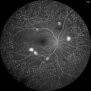
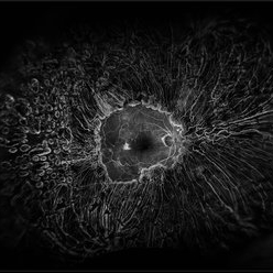
 Active neovascularization in Proliferative Diabetic Retinopathy
Jan 10 2018 by Peter H. Tang, MD, PhD
Fluorescein angiography image from a 46-year-old woman with uncontrolled proliferative diabetic retinopathy shows extensive dye leakage from active neovascularization.
Imaging device: Optos California
Condition/keywords: diabetes, diabetic retinopathy, neovascularization elsewhere (NVE), neovascularization of the disc (NVD), pan-retinal photocoagulation (PRP), proliferative diabetic retinopathy (PDR)
-
 Brach Retinal Artery Occlusion
Oct 2 2013 by Jerald A. Bovino, MD
There is a hollenhorst plaque causing a branch retinal artery occlusion. The patient has scars from prior panretinal laser photocoagulation.
Condition/keywords: branch retinal artery occlusion (BRAO), hollenhorst plaque, pan-retinal photocoagulation (PRP)
-
 Branch Retinal Artery Occlusion
Oct 2 2013 by Jerald A. Bovino, MD
There is a hollenhorst plaque causing a branch retinal artery occlusion. The patient has scars from prior panretinal laser photocoagulation.
Condition/keywords: branch retinal artery occlusion (BRAO), hollenhorst plaque, pan-retinal photocoagulation (PRP)
-
 Color Photo of PDR s/p PRP
Feb 19 2015 by H. Michael Lambert, MD
PDR after PRP.
Condition/keywords: pan-retinal photocoagulation (PRP), proliferative diabetic retinopathy (PDR), regressed
-
 Combined Traction-Rhegmatogenous Retinal Detachment
Apr 8 2019 by Gary R. Cook, MD, FACS
White female with a combined diabetic-related traction-rhegmatogenous retinal detachment; s/p full laser PRP in the past; V.A. = 20/400
Imaging device: Topcon VT-50
Condition/keywords: combined retinal detachment, pan-retinal photocoagulation (PRP), proliferative diabetic retinopathy (PDR)
-
 Completed Pan-Retinal Fill-in Laser Photocoagulation in an Air filled eye at the end of Diabetic Vitrectomy Retina Surgery
Apr 28 2023 by Veer Singh, MS, FVRS, FMRF, FICO (Retina)
Completed Pan-Retinal Fill-in Laser Photocoagulation in an Air filled eye at the end of Diabetic Vitrectomy Retina Surgery
Photographer: Dr. Veer Singh
Condition/keywords: air-filled, pan-retinal photocoagulation (PRP), vitrectomy
 Loading…