Search results (12 results)
-
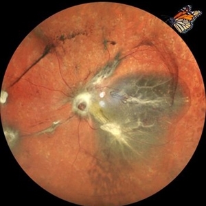
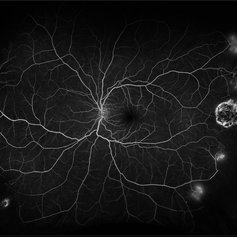
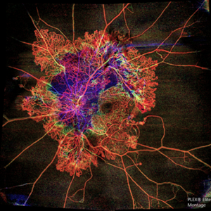 Flame of the Forest
Apr 9 2020 by Daraius N Shroff, MS FMRF FRCS
A 54-year-old man with DM for 15 years. The left eye had a visual acuity of 20/40. Wide field swept source OCTA revealed branching out central neovascular trunk vessels from the disc with terminal loops, along with exuberant proliferation of irregular small-calibre fine new vessels. The patient underwent OCTA guided pan retinal photocoagulation.
Photographer: Anuj Choudhary, Shroff Eye Centre, New Delhi
Imaging device: Zeiss Plex Elite 9000
Condition/keywords: proliferative diabetic retinopathy (PDR)
-
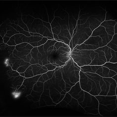 Proliferative Sickle Cell Retinopathy
Feb 1 2023 by Olivia Rainey
Ultra-widefield fluorescein angiography of a 25-year old male with Proliferative Sickle Cell Retinopathy affecting his right eye. Patient stated that he was born with Sickle disease (SC), and has yearly eye exams. He noted no vision concerns over the last year but has typically experienced sickle attacks about 1-2 per year. The physician noted that the fluorescein obtained showed peripheral nonperfusion affecting the patient's nasal and temporal retina as well as neovascularization affecting his left eye more than his right. He recommended pan retinal photocoagulation in his left eye for his temporal and nasal retina, as as well as his right eye following.
Photographer: Olivia Rainey, OCT-C, COA
Imaging device: Optos California
Condition/keywords: early phase, fluorescein angiogram (FA), fluorescein leakage, neovascularization (NV), non-perfusion, proliferative retinopathy, right eye, sickle cell retinopathy, ultra-wide field imaging, ultra-widefield image
-
 Ischemic Central Retinal Vein Occlusion
Jan 24 2019 by Nichole Lewis
76-year-old woman with an ischemic central retinal vein occlusion, severely attenuated and sclerotic vessels and scattered retinal hemorrhages. Vision decrease over 1 year. VA 20/CF. Patient is returning for pan retinal photocoagulation.
Photographer: Nichole Lewis
Imaging device: Optos
Condition/keywords: attenuated vessels, central retinal vein occlusion (CRVO), hemorrhage, ischemic CRVO, sclerotic vessels
-
 Combined Pathology
Oct 26 2024 by rahul saradge
53 year old male patient was presented with a complaints of diminished vision in LE since 1 month. The BCVA in RE was 6/36p and LE was CF 1/2m. Ocular dilated examination showed RE temporal CD with ?CRVO,OIS and OS showed TRD and old Hemi CRVO. Patient was injected with PST tricot followed by PRP laser at an interval of 1 week. Patient improved to BCVA 6/9.
Photographer: Aishwarya Bangar Isha Netralaya Thane
Imaging device: optos
Condition/keywords: choroidal detachment, crvo, ois, optos, pan retinal photocoagulation, tractional retinal detachment
-
 Combined Retinal Detachment With a Butterfly Shaped Configuration
Mar 13 2025 by S. Natarajan, MD, FASRS, FRCS (GLASGOW) , FICO, D.Sc, FELA
A 46 year old female presented to us with diminished vision in both the eyes. Her blood glucose levels were deranged. She had bilateral proliferative diabetic retinopathy and pan retinal photocoagulation was done elsewhere. Left eye showed a combined retinal detachment with fibrovascular proliferation on the disc and along inferior arcade with a convex configuration of retinal detachment. Patient was planned for surgical intervention. The image shows a butterfly like configuration of combined retinal detachment with the subretinal fluid pocket appearing like the wings of the butterfly.
Photographer: ASHWINI SUTAR ADITYA JYOT EYE HOSPITAL
Imaging device: Mirante ( PLEASE SELECT COVER PAGE )
Condition/keywords: retinal detachment with a butterfly shaped
-
 Diabetic Retinopathy
Aug 21 2015 by Andrea Arriola-Lopez, MD MSc
Color fundus photography shows neovascularization of the optic nerve head, macular pre retinal hemorrhage, pan retinal photocoagulation and extreme temporal peripherical retina without PRP.
Photographer: Andrea Elizabeth Arriola L.
Imaging device: OPTOS Dakota
Condition/keywords: diabetes, diabetic retinopathy, neovascularization (NV)
-
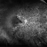
 High risk Proliferative Diabetic Retinopathy treated with Pan Retinal Photocoagulation
Nov 5 2022 by Somnath Chakraborty, MD
A Fundus Photo Montage of 43 year old Asian Male with Type 2 Diabetes Mellitus since 7 years who presented with sudden onset diminition of vision in his Left eye. BCVA OS was 20/200. He was diagnosed to have Pre retinal bleed due to Proliferative Diabetic Retinopathy and was treated with Pan Retinal Photocoagulation. This image shows a large neo-cascular frond at the disc and superior to it with Pre-retinal bleed and Fresh laser marks along
Photographer: Pulak Roy
Condition/keywords: diabetic blindness, diabetic retinopathy vitrectomy study (DRVS), fresh laser burns, laser photocoagulation, preretinal hemorrhage, proliferative diabetic retinopathy (PDR)
-
 Proliferative Diabetic Retinopathy
Jul 12 2018 by Nichole Lewis
68-year-old male with proliferative diabetic retinopathy and capillary nonperfusion. Returning for pan retinal photocoagulation. VA 20/25
Photographer: Nichole Lewis
Condition/keywords: capillary nonperfusion, diabetes, non-perfusion, pan-retinal photocoagulation (PRP), proliferative diabetic retinopathy (PDR)
-
 Proliferative Diabetic Retinopathy S/P Pan Retinal Photocoagulation
Mar 4 2025 by Prithvi Chandrakanth
A 52-year-old female patient presented with complaints of diminishing vision, compounded by uncontrolled diabetes mellitus. Her Fundus examination revealed proliferative diabetic retinopathy, characterized by neovascularization of the disc and elsewhere, and sclerosed vessels. To address this, Pan Retinal Photocoagulation was performed, and the condition stabilized, halting the progression of the disease.
Photographer: DR PRITHVI CHANDRAKANTH, DR CHANDRAKANTH NETHRALAYA, KOZHIKODE, KERALA, INDIA
Imaging device: EIDON
Condition/keywords: Diabetic Retinopathy, Neovascularisation at the Disc (NVD), neovascularization of the disc (NVD), NVD, pan-retinal photocoagulation (PRP), PDR, PDR with NVE (periphery), PRP
-
 Proliferative Sickle Cell Retinopathy
Feb 1 2023 by Olivia Rainey
Ultra-widefield fluorescein angiography of a 25-year old male with Proliferative Sickle Cell Retinopathy affecting his left eye. Patient stated that he was born with Sickle disease (SC), and has yearly eye exams. He noted no vision concerns over the last year but has typically experienced sickle attacks about 1-2 per year. The physician noted that the fluorescein obtained showed peripheral nonperfusion affecting the patient's nasal and temporal retina as well as neovascularization affecting his left eye more than his right. He recommended pan retinal photocoagulation in his left eye for his temporal and nasal retina, as as well as his right eye following.
Photographer: Olivia Rainey, OCT-C, COA
Imaging device: Optos California
Condition/keywords: early phase, fluorescein angiogram (FA), fluorescein leakage, left eye, neovascularization (NV), proliferative retinopathy, sickle cell retinopathy, ultra-wide field imaging, ultra-widefield image
-
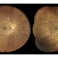
 PRP Marks
Apr 26 2021 by Priya Rasipuram Chandrasekaran, MBBS, DO, DNB, FRCS
This is the fundus photo montage of both eyes of a patient showing pan retinal photocoagulation marks. Theses marks can be confused with gyrate atrophy, cobble stone degeneration and myopic degeneration.
Condition/keywords: pan-retinal photocoagulation (PRP)
-
 Subhyaloid Hemorrhage in the Setting of PDR, Now s/p PRP Laser
Jan 22 2021 by Vishak J. John, MD
60-year-old man with proliferative diabetic retinopathy presented with central loss of vision. He underwent pan retinal photocoagulation.
Photographer: Danielle Lombardo, Vistar Eye Center
Imaging device: Optos
Condition/keywords: proliferative diabetic retinopathy (PDR)
 Loading…