Search results (16 results)
-
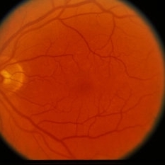
 Inactive Toxoplasmosis
Nov 9 2012 by Norman Byer
This 28-year-old man had inactive toxoplasmosis and presented with acute symptoms caused by this tractional retinal tear adjacent to a retinochorodial scar. He also had an acute posterior vitreous detachment which had torn this retinal operculum completely free. The next slide shows the same lesion. Note the early rolled edge on the left side of the tear.
Condition/keywords: acute posterior vitreous detachment, inactive toxoplasmosis, operculum, rolled edges of retina, tractional retinal tear
-
 Asymptomatic Tractional Tear
Nov 9 2012 by Norman Byer
This 38-year-old man was found to have this asymptomatic tractional tear in which the vitreoretinal traction had completely avulsed this tiny fragment of retina as a free operculum. Note how the examination and also the photography of this tiny lesion is made easier by scleral indentation.
Condition/keywords: asymptomatic, free operculum, scleral indentation, vitreoretinal traction
-
 Inactive Toxoplasmosis
Nov 9 2012 by Norman Byer
This is the same case as in the previous photograph showing the very large free operculum torn from the retina.
Condition/keywords: acute posterior vitreous detachment, free operculum, inactive toxoplasmosis, tractional retinal tear
-
 Laser Photocoagulation
Nov 9 2012 by Norman Byer
This is the same lesion 18 days following photocoagulation. The continuing vitreoretinal traction has now torn the retinal flap completely away from the retina and the resulting free operculum may be seen out of focus in the lower part of the photograph. The retinal tear is now easily visible with only a tiny remaining nubbin of the original flap seen above with a small hemorrhage.
Condition/keywords: free operculum, laser photocoagulation, retinal tear, vitreoretinal traction
-
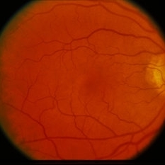
 Retinal Detachment
Nov 9 2012 by Norman Byer
The retinal detachment in this 52-year-old man was brought on by this oval tear which has no flap or free operculum. This is however still a tractional tear caused by the retina pulling away from a corneoretinal adhesion, which is marked by the yellow spots seen through the tear. Actually, therefore, the operculum is behind the retina and attached to the pigment epithelial layer.
Condition/keywords: corneoretinal adhesion, operculum, tractional retinal tear
-
 Slide 8-1
Mar 4 2019 by Lancaster Course in Ophthalmology
Left: Gross appearance of a funnel-shaped detachment of the vitreous. It remains attached at its base and at the optic nerve head. Center and right: Appearance of ring-shaped vitreous condensation, formerly attached at the optic nerve head. Such condensations can account for the visual symptom of floaters. (E.P. No. 21811)
Condition/keywords: detachment, operculum, vitreous
-
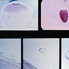 Slide 8-6
Mar 4 2019 by Lancaster Course in Ophthalmology
Gross and microscopic appearance of a retinal hole. Vitreous tractim has resulted in the hole and detachment of the operculum. Upper right shows the plug of retinal tissue (arrow) attached to the posterior aspect of the detached vitreous. Lower left shows the rounded posterior margin of the retinal hole and the small area of associated retinal detachment. Lower middle and right views are of sections through the detached retinal operculum. (E.P. No. 35238)
Condition/keywords: operculum, retinal hole, retinal tissue
-
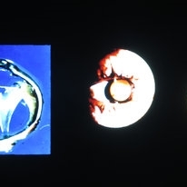
 Stage 3 Macular Hole With Operculum
Sep 25 2018 by samarth mishra
Stage 3 macular hole with operculum.
Photographer: Aditya Birla Sankara Nethralaya, Kolkata, West Bengal , India and Sankara Nethralaya, chennai , India
Condition/keywords: full thickness macular hole, macular hole, optical coherence tomography (OCT)
-
 Sudden Posterior Vitreous Detachment
Nov 9 2012 by Norman Byer
This is the appearance of the previous lesion three weeks following prophylactic cryotherapy. Continuing vitreal retinal traction has a now torn the flap completely free from the retina. The whitish cystic retinal tuft can be discerned on the upper part of the free operculum. Along the lower half of the operculum superimposed over the dark shadow of the scleral indentation one may observe numerous, delicate, vitreous fibrils actually attaching to the operculum.
Condition/keywords: cystic retinal tuft, free operculum, prophylactic cyrotherapy, retinal flap, scleral indentation, vitreoretinal traction, vitreous fibrils
-
 Tractional Retinal Tear
Nov 9 2012 by Norman Byer
This is the same lesion and shows the free operculum in better focus. This is an unusual location for a tractional retinal tear, and the increased mobility of the detached vitreous in the posterior part of the eye may have been a factor leading to the complete rupture of this retinal flap.
Condition/keywords: detached vitreous, free operculum, tractional retinal tear
-
 White Retinal Tuft
Nov 9 2012 by Norman Byer
After six years, the previous lesion looked like this. The former flap has been completely avulsed and is now a free operculum. The white zone around the tear represents the small area of detachment and subretinal fluid. It is still asymptomatic and does not require treatment.
Condition/keywords: does not require treatment, free operculum, operculated retinal hole, subretinal fluid, white retinal tuft
-
 Yellow Spot Pseudo-Operculum
Oct 31 2014 by David Callanan, MD
Male patient, yellow spot pseudo-operculum.
Condition/keywords: operculum
-
 Yellow Spot Pseudo-Operculum
Oct 31 2014 by David Callanan, MD
Male patient, yellow spot pseudo-operculum.
Condition/keywords: operculum
-
 Yellow Spot Pseudo-Operculum
Oct 31 2014 by David Callanan, MD
Male patient, yellow spot pseudo-operculum.
Condition/keywords: operculum
-
 Yellow Spot Pseudo-Operculum
Oct 31 2014 by David Callanan, MD
Male patient, yellow spot pseudo-operculum.
Condition/keywords: operculum
-
 Yellow Spot Pseudo-Operculum
Oct 31 2014 by David Callanan, MD
Male patient, yellow spot pseudo-operculum.
Condition/keywords: operculum
 Loading…