Search results (111 results)
-
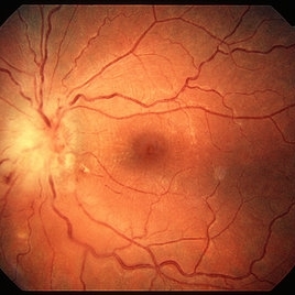
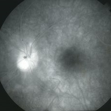
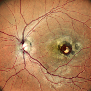
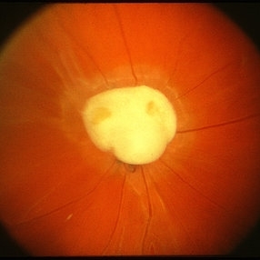 Toxocara Granuloma
Feb 25 2013 by Henry J. Kaplan, MD
Toxocara granuloma of the optic nerve head.
Condition/keywords: ocular toxoplasmosis, toxocara granuloma, toxocariasis
-
 Childhood Acquired Ocular Toxoplasmosis
Sep 13 2023 by Deepak Bhojwani, MS
Fundus image of a 16 year old boy diaagnosed with Ocular Toxoplasmosis since the age of 10 years showing the classic toxo chorioretinitis scar on the posterior pole. Luckily the scar is loacted juxtatemporal to fovea on OCT and so the boy has good vision of 20/30.
Photographer: DR DEEPAK BHOJWANI
Imaging device: OPTCAL COHERENCE TOMOGRAPHY
Condition/keywords: posterior uveitis, toxo chorioretinitis
-
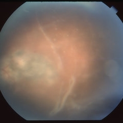
 Thickening of the Posterior Hyaloid
Dec 12 2020 by Anyssa Montenegro
Color fundus photograph of the right eye of a 36-year-old man showing thickening of the posterior hyaloid associated with an epiretinal membrane due to ocular toxoplasmosis.
Photographer: Anyssa Montenegro, Centro Brasileiro da Visão, Brasília-DF, Brazil
Condition/keywords: epiretinal membrane (ERM), ocular toxoplasmosis, thickening of the posterior hyaloid
-
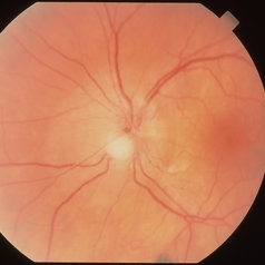
 Toxocara Granuloma
Feb 25 2013 by Henry J. Kaplan, MD
Toxocara granuloma superotemporal to the fovea.
Condition/keywords: ocular toxoplasmosis, toxocara granuloma, toxocariasis
-
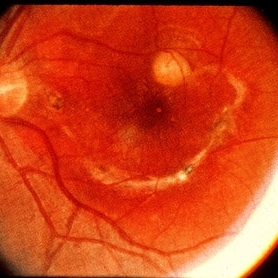
 Toxoplasma chorioretinitis 1
Jan 11 2013 by Alex P. Hunyor, MD
Toxoplasmosis 1 - chorioretinal scar from previous toxoplasma chorioretinitis. See image 2 - recurrent todo adjacent to this scar
Condition/keywords: inactive toxoplasmosis, ocular toxoplasmosis, toxoplasmosis, toxoplasmosis retinitis
-
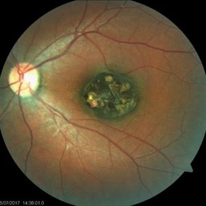
 Toxoplasmosis
Jun 3 2017 by Gabriel Costa Andrade, PhD
Fundus photograph of an 14-year-old boy with multiple chorioretinal scars secondary to toxoplasmosis.
Photographer: Gabriel Costa de Andrade
Imaging device: Optos® California
Condition/keywords: congenital toxoplasmosis, ocular toxoplasmosis, toxoplasmosis chorioretinitis, toxoplasmosis uveitis
-
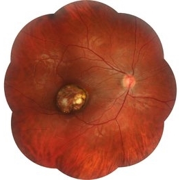
 Toxocara Granuloma
Feb 25 2013 by Henry J. Kaplan, MD
Toxocara granuloma in the midperiphery of the retina.
Condition/keywords: ocular toxoplasmosis, toxocara granuloma, toxocariasis
-
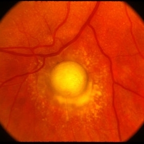
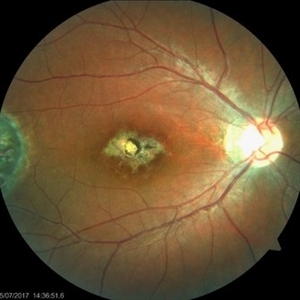
 Toxoplasma Neuroretinitis (Jensen`s Disease)
Feb 25 2013 by Henry J. Kaplan, MD
Toxoplasma neuroretinitis in the left eye of a patient with macular star formation, retinitis adjacent to the optic nerve head with disc swelling.
Condition/keywords: Jensen disease, ocular toxoplasmosis, toxoplasmosis
-
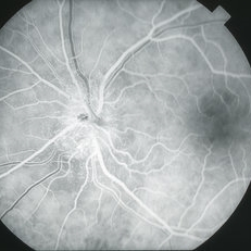
 Ocular Toxoplasmosis Scar, Fluorescein Angiogram
Aug 23 2012 by Gerardo Garcia-Aguirre, MD
Fluorescein angiogram showing a large hypofluorescent round lesion with well-defined borders, where the fluorescence of the choroidal vessels is observed.
Photographer: Noemí Hernández, Asociación para Evitar la Ceguera en México
Imaging device: Zeiss FF4
Condition/keywords: toxoplasmosis
-
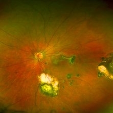
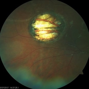
 'Wet Snow on Grapevines'
Apr 8 2019 by Gary R. Cook, MD, FACS
Fundus photograph of inflammatory deposits on vitreous fibrils, known as "Wet snow on grapevines" in a case of recurrent ocular toxoplasmosis.
Imaging device: Topcon VT-50
Condition/keywords: ocular toxoplasmosis, vitritis, wet snow on grapevines
-
 Aborted Arteriolitis
Feb 15 2013 by From the Collections of Thomas M. Aaberg, MD and Thomas M. Aaberg Jr., MD
Fundus photograph showing activated toxoplasma retinochoroiditis with active retinal whitening adjacent to a hyperpigmented scar in the superonasal mid-periophery.
Condition/keywords: ocular toxoplasmosis
-
---thumb.jpg/image-square;max$300,300.ImageHandler) Aborted Arteriolitis - diffuse hyper-permeability and staining of the infectious retinal lesion
Aborted Arteriolitis - diffuse hyper-permeability and staining of the infectious retinal lesion
Feb 15 2013 by From the Collections of Thomas M. Aaberg, MD and Thomas M. Aaberg Jr., MD
Fluorescein angiogram corresponding to slide titled Aborted Arteriolitis showing diffuse hyper-permeability and staining of the infectious retinal lesion.
Condition/keywords: ocular toxoplasmosis
-
---thumb.jpg/image-square;max$300,300.ImageHandler) Active retinitis consistent with ocular toxoplasmosi
Active retinitis consistent with ocular toxoplasmosi
Feb 15 2013 by From the Collections of Thomas M. Aaberg, MD and Thomas M. Aaberg Jr., MD
Fundus photograph showing active retinitis consistent with ocular toxoplasmosis.
Condition/keywords: ocular toxoplasmosis
-
 Acute Toxoplasmosis in AIDS
Apr 8 2019 by Gary R. Cook, MD, FACS
Left eye of a white male with AIDS and an optic neuritis secondary to ocular toxoplasmosis infection. The patient had no pre-existing chorioretinal scars secondary to Toxo. An edematous optic nerve with a focus of active retinitis inferonasally, small surface hemorrhage above it, and surrounding peripapillary edema is visible.
Imaging device: Topcon VT-50
Condition/keywords: AIDS, ocular toxoplasmosis, optic neuritis, toxoplasmosis
-
 AIDS/Toxoplasmosis
Apr 8 2019 by Gary R. Cook, MD, FACS
Laminar venous phase FA of white male with AIDS and optic neuritis secondary to ocular toxoplasmosis OS showing dilated capillaries on the surface of the optic nerve and relative hypofluoescence due to peripapillary edema.
Imaging device: Topcon VT-50
Condition/keywords: AIDS, ocular toxoplasmosis, optic neuritis, toxoplasmosis
-
 AIDS/Toxoplasmosis
Apr 8 2019 by Gary R. Cook, MD, FACS
Late-phase frame of FA of white male with AIDS and optic neuritis secondary to ocular toxoplasmosis OS showing diffuse late staining of optic nerve.
Imaging device: Topcon VT-50
Condition/keywords: AIDS, ocular toxoplasmosis, optic neuritis, toxoplasmosis
-
 Bilateral Ocular Toxoplasmosis
Oct 30 2022 by Vaibhavi Noticewala, M S Ophthalmology, FVRS
Bilateral Ocular Toxoplasmosis
Photographer: Optom Priyal Mistry
Condition/keywords: toxoplasmosis
-
 Bilateral Ocular Toxoplasmosis
Oct 30 2022 by Vaibhavi Noticewala, M S Ophthalmology, FVRS
Bilateral Ocular Toxoplasmosis
Photographer: Optom Priyal Mistry
Condition/keywords: toxoplasmosis
-
 Childhood Acquired Ocular Toxoplasmosis
Sep 13 2023 by Deepak Bhojwani, MS
Fundus image of a 16 year old boy diaagnosed with Childhood Acquired Ocular Toxoplasmosis since the age of 10 years showing the classic toxo chorioretinitis scar on the posterior pole complicated by Choroidal neovascularisation over the macula. OCT documenting complete foveal atrophy.
Photographer: DR DEEPAK BHOJWANI
Imaging device: OPTICAL COHEERENCE TOMOGRAPHY
Condition/keywords: choroidal neovascularization (CNV), toxo chorioretinitis
-
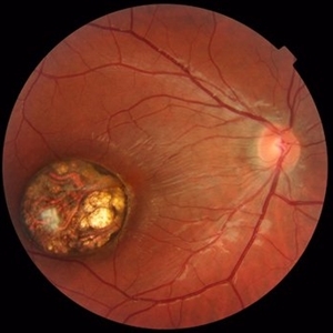
 Choroidal Neovascular Membrane in a case of Reactivated Ocular Toxoplasmosis
Dec 29 2022 by Vaidehi Sathaye
Fundus Photograph of LE of a 34 year old female with a CNVM secondary to Reactivated Ocular Toxoplasmosis
Photographer: Dr. Vaidehi Sathaye
Imaging device: Mirante
Condition/keywords: choroidal neovascular membrane (CNVM), ocular toxoplasmosis
-
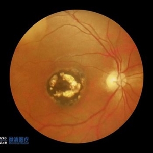
 Congenital Toxoplasmosis
Jul 22 2017 by Akif Erol
Color fundus photograph of the left eye of an 18-year-old girl with decreased vision due to a large chorioretinal scar involving the macula. The lesion is typical for a congenital ocular toxoplasmosis
Photographer: Mehmet Akif Erol, Afyon Kocatepe University Ophthalmology Clinic
Condition/keywords: color fundus photograph, congenital toxoplasmosis
-
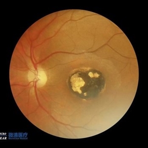
 Congenital Toxoplasmosis
Jul 22 2017 by Akif Erol
Color fundus photograph of the right eye of an 18-year-old girl with decreased vision due to a large chorioretinal scar involving the macula. The lesion is typical for a congenital ocular toxoplasmosis
Photographer: Mehmet Akif Erol, Afyon Kocatepe University Ophthalmology Clinic
Condition/keywords: color fundus photograph, congenital toxoplasmosis
-
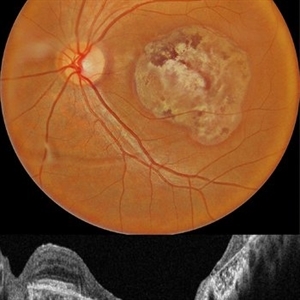
 Congenital Toxoplasmosis
Oct 10 2015 by Hamid Ahmadieh, MD
Wide-field color fundus photograph of the right eye of a 15 -year-old boy with decreased vision due to a large chorioretinal scar involving the macula . The lesion is typical for a congenital ocular toxoplasmosis .
Photographer: Solmaz Shahmohammad, Negah Eye Center, Tehran, Iran
Condition/keywords: color fundus photograph, congenital toxoplasmosis
-
 Congenital Toxoplasmosis
Oct 10 2015 by Hamid Ahmadieh, MD
Color fundus photograph of the right eye of a 15 -year-old boy with decreased vision due to a large chorioretinal scar involving the macula . The lesion is typical for a congenital ocular toxoplasmosis .
Photographer: Solmaz Shahmohammad, Negah Eye Center, Tehran, Iran
Condition/keywords: color fundus photograph, congenital toxoplasmosis
 Loading…