Search results (5 results)
-
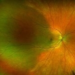 Isolated Choroidal Melanocytosis
Oct 27 2019 by John S. King, MD
23-year-old white female consulted for a large pigmented choroidal lesion in the right eye. Healthy, no history of glaucoma, 20/20 OU without scleral pigment changes; large, flat pigmented choroidal lesion that is sectoral (temporally) and pigment appears to spare the major choroidal vessels. On OCT (not shown) there is mildly increased choroidal thickening in the area of the lesion.
Photographer: Shelly Blair
Imaging device: Optos CA
Condition/keywords: choroidal melanocytosis, ocular melanocytosis
-
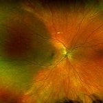 Isolated Choroidal Melanocytosis - Montage
Oct 27 2019 by John S. King, MD
23-year-old white female consulted for a large pigmented choroidal lesion in the right eye. Healthy, no history of glaucoma, 20/20 OU without scleral pigment changes; large, flat pigmented choroidal lesion that is sectoral (temporally) and pigment appears to spare the major choroidal vessels. On OCT (not shown) there is mildly increased choroidal thickening in the area of the lesion.
Photographer: Shelly Blair
Imaging device: Optos CA
Condition/keywords: choroidal melanocytosis, ocular melanocytosis
-
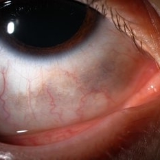 Ocular Melanocytosis Scleral Pigmentation
Jul 9 2014 by Susanna S. Park, MD, PhD
Slit lamp photograph of a 12-year-old boy with ocular melanocytosis showing scleral and episcleral pigmentation. The other eye is blue.
Photographer: Ellen Redenbo
Condition/keywords: melanocytoma
-
 Ocular Melanocytosis w/Treated Melanoma
Jan 27 2025 by Virginia Gebhart
74 year female with ocular melanocytosis. Stable, regressed treated tumor s/p brachytherapy (2020) and deeply pigmented fundus OS. Limited VA due to radiation neuropathy. BCVA 20/150 (ecc)
Photographer: Virginia Gebhart, Retina Consultants of Carolina
Imaging device: Optos California
Condition/keywords: brachytherapy, melanoma, melanosis, ocular melanocytosis
-
 Sectoral Ocular Melanocytosis
Jan 17 2025 by Virginia Gebhart
67 year old female with congenital sectoral ocular melanocytosis. Pigmentation on nasal sclera and nasal iris of right eye, as well as deep pigmentation nasally of fundus. Will continue close observation
Photographer: Virginia Gebhart
Imaging device: Topcon 50DX/Samsung Galaxy
Condition/keywords: choroidal melanocytosis, heterochromia, ocular melanocytosis, Oculodermal Melanocytosis
 Loading…