Search results (33 results)
-
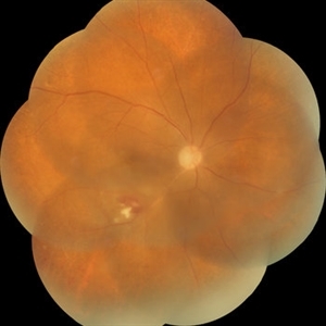
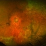
 Superior Hemi-Central Retinal Artery Occlusion
Apr 24 2024 by Mosab Salah
Fundus photograph -inverted view- taken by smartphone fundus photography, of a young man with sudden onset altitudinal field defect, a Superior Hemi-Central Retinal Artery Occlusion noted.
Photographer: Dr Mosab Salah, The Islamic Hospital, Amman, Jordan
Imaging device: smartphone fundus photography and 30 D Lens
Condition/keywords: arterial occlusion, branch retinal artery occlusion (BRAO), BRAO, CRAO, Hemi-Central Retinal Artery Occlusion (CRAO), occlusive vasculitis, smartphone fundus photography
-
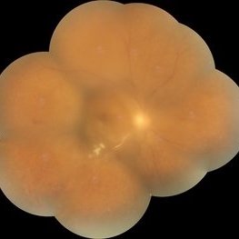
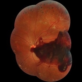 Subhyaloid Hemorrhage
Jul 7 2015 by Hamid Ahmadieh, MD
Color fundus photograph of the left eye of a 25-year-old woman with severe subhyaloid hemorrhage due to an advanced vasoproliferative vitreoretinopathy secondary to a severe idiopathic occlusive retinal vasculitis.
Photographer: Soulmaz Shahmohammad, Negah Eye Center, Tehran, Iran
Condition/keywords: color fundus photograph, occlusive vasculitis, subhyaloid hemorrhage
-
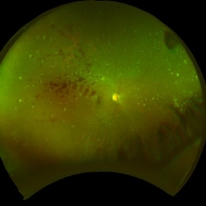
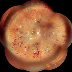 Atypical Tubercular Peripheral Occlusive Retinal Vasculitis
Jun 21 2024 by Tejaswita Verma
Fundus montage of the right eye of a 27 year old male with macula threatening occlusive vasculitis showing hemorrhages in inferior, temporal quadrant with vascular sheathing. The patient was Mantoux positive (20 mm induration) and IGRA (TB-GOLD)positive and started on oral steroids. The case was atypical due to no vitritis at presentation which is unusual of tuberculosis. Behcet's disease was ruled out as there was no panuveitis like picture.
Photographer: DR. TEJASWITA VERMA
Imaging device: MIRANTE
Condition/keywords: occlusive vasculitis, ocular tuberculosis
-
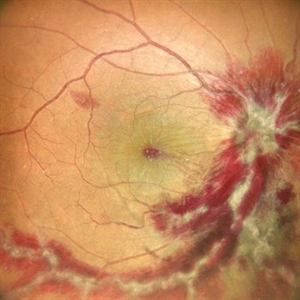
 Behcet's Disease
Nov 25 2012 by Mallika Goyal, MD
Fundus photograph of right eye of a 23-year-old gentleman with Behcet's Disease shows occlusive retinal vasculitis with optic disc pallor and macular ischaemia. Other eye has similar appearance with no light perception.
Photographer: Mallika Goyal, MD, Apollo Health City, Hyderabad, India
Condition/keywords: occlusive vasculitis
-
---thumb.JPG/image-square;max$300,300.ImageHandler) Behcet's Disease
Behcet's Disease
Nov 25 2012 by Mallika Goyal, MD
Fundus photograph of right eye of a 23-year-old gentleman with Behcet's Disease shows occlusive retinal vasculitis with optic disc pallor and macular ischaemia. Other eye has similar appearance with no light perception.
Photographer: Mallika Goyal, MD, Apollo Health City, Hyderabad, India
Condition/keywords: macular ischemia, occlusive vasculitis
-
 Behcet's Disease
Nov 25 2012 by Mallika Goyal, MD
Fundus photograph of left eye of a 23-year-old gentleman with Behcet's Disease shows occlusive retinal vasculitis with optic disc pallor and macular ischemia. This eye has no light perception; other eye has similar fundus appearance.
Photographer: Mallika Goyal, MD, Apollo Health City, Hyderabad, India
Condition/keywords: macular ischemia, occlusive vasculitis, optic disc pallor
-
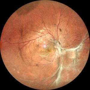 Extramacular TRD in Idiopathic Occlusive Vasculitis
Dec 5 2024 by Tejaswita Verma
Fundus photo showing extramacular TRD in a 16 year old boy with idiopathic occlusive vasculitis secondary to presumed IOTB. History of taking ATT for 6 months , Mantoux positive previously. Vision was 6/6P,other eye had funnel RD .
Photographer: DR. TEJASWITA VERMA
Imaging device: MIRANTE
Condition/keywords: tractional retinal detachment, vasculitis
-
 Fundus Photo Montage showing Occlusive Vasculitis from Brolucizumab: 1-week post treatment with Steroids
Jan 21 2022 by Somnath Chakraborty, MD
Right eye of a 62-year old lady with inferotemporal Branch Retinal Vein Occlusion, treated with single dose of "off-label" brolucizumab. She developed Occlusive Vasculitis 9 weeks post injection. We treated her with topical and systemic steroids. This is her Fundus Photo Montage after 1 week of therapy. BCVA OD 20/200.
Photographer: Pulak Roy
Condition/keywords: branch retinal vein occlusion (BRVO), Brolucizumab, occlusive vasculitis
-
 Fundus Photo Montage showing Occlusive Vasculitis from Brolucizumab: 3 weeks Post treatment with Steroids
Jan 21 2022 by Somnath Chakraborty, MD
Right eye of a 62-year-old lady with inferotemporal Branch Retinal Vein Occlusion, treated with single dose of "off-label" brolucizumab. She developed Occlusive Vasculitis 9 weeks post injection. We treated her with topical and systemic steroids. This is her Fundus Photo Montage, 3 weeks after therapy. BCVA OD 20/80.
Photographer: Pulak Roy
Condition/keywords: branch retinal vein occlusion (BRVO), Brolucizumab, occlusive vasculitis
-
 Fundus Photo Montage showing Occlusive Vascultis from Brolucizumab
Jan 21 2022 by Somnath Chakraborty, MD
Right eye of a 62-year-old lady with Inferotemporal Branch Retinal Vein Occlusion, treated with single dose of "off-label" brolucizumab. She developed Occlusive Vasculitis 9 weeks post injection. This is her Fundus Photo Montage at that time, showing evidence of Occlusive Vasculitis with moderate grade vitritis. BCVA OD 20/400.
Photographer: Pulak Roy
Condition/keywords: branch retinal vein occlusion (BRVO), Brolucizumab, occlusive vasculitis, vitritis
-
 Lupus Hemorrhagic Occlusive Vasculitis
Apr 23 2018 by Frank Chin
Fundus photograph of the right eye of a 24-year-old woman with history of systemic lupus erythematosus who presented with decreased visual acuity for 2-3 days found to have lupus hemorrhagic occlusive vasculitis with mild disc elevation, diffuse punctate cotton wool spots and dot blot hemorrhages, and a hemorrhage occlusive vasculitis along the superior branch of the superotemporal arcade involving the artery and vein.
Photographer: Frank Chin, MD, George Washington University
Imaging device: Optos 200Tx
Condition/keywords: blot hemorrhages, cotton wool spots, occlusive vasculitis, systemic lupus erythematosus (SLE) vasculitis
-
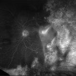
 Occlusive Retinal Vasculitis
Dec 17 2020 by Somnath Chakraborty, MD
Fundus photo montage of left eye of a 45-year-old male showing a large neovascular frond secondary to peripheral occlusive vasculitis.
Photographer: Pulak Roy
Condition/keywords: neovascularization elsewhere (NVE), peripheral retinal vasculitis, retinal vasculitis
-
 Occlusive Vasculitis
Mar 9 2013 by Gabriela Lopezcarasa Hernandez, MD
A 36-year-old women with panuveitis and bilateral occlusive vasculitis.
Photographer: Azucena Rios Macula Retina Consultores Mexico
Imaging device: heidelberg Spectralis
Condition/keywords: occlusive vasculitis
-
 Occlusive Vasculitis
Mar 9 2013 by Gabriela Lopezcarasa Hernandez, MD
A 30-year-old woman with decrease in visual acuity in both eyes secondary to an autoimmune response.
Photographer: Araceli Rojas Arriaga, Hospital Angeles Lomas, Mexico
Imaging device: Zeiss FF4
Condition/keywords: occlusive vasculitis
-
 Occlusive Vasculitis
Mar 9 2013 by Gabriela Lopezcarasa Hernandez, MD
A 30-year-old woman with decrease in visual acuity in both eyes secondary to an autoimmune response.
Photographer: araceli Rojas Arriaga, Hospital Angeles Lomas, Mexico
Imaging device: Zeiss FF4
Condition/keywords: occlusive vasculitis
-
 Occlusive Vasculitis
Mar 9 2013 by Gabriela Lopezcarasa Hernandez, MD
A 30-year-old woman with decrease in visual acuity in both eyes secondary to an autoimmune response.
Photographer: Araceli Rojas Arriaga, Hospital Angeles Lomas, Mexico
Imaging device: Zeiss FF4
Condition/keywords: occlusive vasculitis
-
 Occlusive Vasculitis
Mar 9 2013 by Gabriela Lopezcarasa Hernandez, MD
30-year-old woman with decrease in visual acuity in both eyes secondary to an autoimmune response.
Photographer: Araceli Rojas Arriaga, Hospital Angeles Lomas, Mexico
Imaging device: zeISS FF4
Condition/keywords: occlusive vasculitis
-
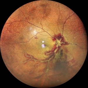
 Occlusive Vasculitis
Jan 28 2023 by Anjana Mirajkar, MS Ophthalmology
Central color image of RE of a 40 year old female a case of occlusive retinal vasculitis
Photographer: Dr. Anjana Mirajkar -Retina Foundation, Ahmedabad
Condition/keywords: occlusive retinal vasculitis
-
 Occlusive Vasculitis
Jan 28 2023 by Anjana Mirajkar, MS Ophthalmology
Widefield color image of RE of a 40 year old female a case of occlusive retinal vasculitis.
Photographer: Dr. Anjana Mirajkar -Retina Foundation, Ahmedabad
Condition/keywords: occlusive retinal vasculitis
-
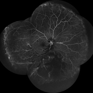 Occlusive Vasculitis
Jan 28 2023 by Anjana Mirajkar, MS Ophthalmology
Wide field FA image of RE of a 40 year old female a case of occlusive retinal vasculitis.
Photographer: Dr. Anjana Mirajkar -Retina Foundation, Ahmedabad
Condition/keywords: occlusive retinal vasculitis
-
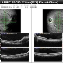 Occlusive Vasculitis
Jan 28 2023 by Anjana Mirajkar, MS Ophthalmology
OCT image of BE of a 40 year old female a case of occlusive retinal vasculitis.
Photographer: Dr. Anjana Mirajkar -Retina Foundation, Ahmedabad
Condition/keywords: occlusive retinal vasculitis
-
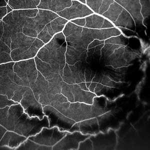 Occlusive Vasculitis
Jan 28 2023 by Anjana Mirajkar, MS Ophthalmology
Central FA picture of a 40 year old female a case of occlusive retinal vasculitis.
Photographer: Dr. Anjana Mirajkar -Retina Foundation, Ahmedabad.
Condition/keywords: occlusive retinal vasculitis
-
 Progressive Outer Retinal Necrosis
Nov 30 2018 by Nichole Lewis
Fluorescein angiogram of an 86-year-old male with progressive outer retinal necrosis and chronic cystoid macular edema. This patient has occlusive vasculitis with non-perfusion, significant retinitis, retinal whitening and intra-retinal hemorrhages. Patient is immunosupressed with a history of kidney transplantation. VA 20/60. Patient was treated with intravitreal foscarnet and admitted to the hospital for an infectious disease and transplant team consultation.
Photographer: Nichole Lewis
Condition/keywords: cystoid macular edema (CME), intraretinal hemorrhage, non-perfusion, occlusive vasculitis, progressive outer retinal necrosis (PORN), retinal whitening, retinitis
-
 Progressive Outer Retinal Necrosis
Nov 30 2018 by Nichole Lewis
Fluorescein angiogram of an 86-year-old male with progressive outer retinal necrosis and chronic cystoid macular edema. This patient has occlusive vasculitis with non-perfusion, significant retinitis, retinal whitening and intra-retinal hemorrhages. Patient is immunosupressed with a history of kidney transplantation. VA 20/60. Patient was treated with intravitreal foscarnet and admitted to the hospital for an infectious disease and transplant team consultation.
Photographer: Nichole Lewis
Condition/keywords: cystoid macular edema (CME), intragel hemorrhage, non-perfusion, occlusive vasculitis, progressive outer retinal necrosis (PORN), retinal whitening, retinitis
-
 Progressive Outer Retinal Necrosis
Nov 5 2019 by Nichole Lewis
86-year-old male with progressive outer retinal necrosis, significant retinitis, retinal whitening, intraretinal hemorrhages and peripheral rpe changes. FA showed occlusive vasculitis with non-perfusion. Patient is immuno-suppressed with a history of renal transplant. VA 20/60.
Photographer: Nichole Lewis
Imaging device: Optos
Condition/keywords: intraretinal hemorrhage, occlusive vasculitis, progressive outer retinal necrosis (PORN), retinal pigment epithelium (RPE) changes, retinal whitening, retinitis
 Loading…