Search results (9 results)
-
 Serpigenous Choroidopathy in a 68-Year-Old Male
Feb 15 2013 by Roy Schwartz, MD
A 68-year-old healthy male presented with a few years of decreased vision bilaterally. Visual acuity in OD was 1/36 and in OS 20/40. Anterior segments were normal except for bilateral mild nuclear sclerosis and pseudoexfoliation in OS. In the fundus of OD a large atrophy with pigmentary scars were seen in the macula and nasally to the optic disc while OS presented with the same clinical picture but an island of normal appearing retina was seen in the fovea. On fluorscein angiography no leakage was shown. A diagnosis of Serpigenous choroidopathy was made.
Photographer: Galit Yair-Pur
Condition/keywords: macula serpiginous choroidopathy, serpiginous choroiditis
-
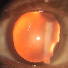
 Congenital Nuclear Cataract
Jul 5 2024 by Zach Seim
This is a slit-lamp photograph of a 10 year old female with a congenital nuclear cataract OD. Patient presented with VA Dsc 20/200. Patient was counseled on surgical options.
Photographer: Zach Seim
Imaging device: Slit Lamp Photography on Samsung Galaxy 7
Condition/keywords: cataract, congenital cataract, nuclear sclerosis, right eye, slit lamp photo
-
---thumb.jpg/image-square;max$300,300.ImageHandler) Hypertensive Retinopathy
Hypertensive Retinopathy
Oct 15 2013 by Maurice F. Rabb
The patient is a 61 year old female who first noted decreased vision in OS one year ago. The right eye is asymptomatic. The patient has systemic hypertension. There is a questionable history of angina. She has low back pain and leg pains of a non-specific nature. Uncorrected vision is OD was 20/25-, not improvable, and in OS 20/400, not improvable. Blood pressure recorded in the office was 230/105. There was mild nuclear sclerosis. Both fundi are characterized by multiple vascular abnormalities consistent with nerve fibers infarcts, hemorrhages (both deep and superficial), and occasional microaneurysms. In OS, there is a large subretinal and preretinal hemorrhage in the macula. This is surrounded by a wreath of outer retinal exudates.
Condition/keywords: hypertensive retinopathy
-
 Lens Coloboma
Oct 17 2018 by Mehul A Shah
35-year-old male presented with diminished vision. On examination he was having nuclear sclerosis and lens coloboma.
Photographer: MEHUL SHAH
Condition/keywords: coloboma
-
 Slide 10-1
Feb 26 2019 by Lancaster Course in Ophthalmology
Senile cataract (PAS x 16). Section shows liquefaction of the cortex under a deeply staining posterior capsule and a dense, lighter staining nuclear sclerosis. (Scheie Eye Institute, No. 4042.)
Condition/keywords: cataract
 Loading…