Search results (50 results)
-
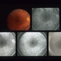
 Coats' Disease - Stage 3A
Aug 21 2019 by Victor M Villegas, MD
Coats' Disease - stage 3A.
Condition/keywords: abnormal retina, Coats' disease, diffuse lipid exudate, edema, foveal hard exudates, pediatic retina, retcam, retinal angioma
-
 Sunset Glow Fundus
May 15 2022 by Manuel Ángel Alcántara Delgado, MD
Optomap ultra-widefield retinal imaging of an 35-year-old woman showed sunset glow fundus, multiple nummular chorioretinal atrophic lesions, macular subretinal fibrosis and pigment clumping in chronic recurrent stage of Vogt-Koyanagi-Harada disease.
Photographer: Manuel Ángel Alcántara Delgado. Conde de Valenciana.
Condition/keywords: abnormal retina, benign pigmented lesions, pigment clumps, retinal fibrosis, uveitis, Vogt-Koyanagi-Harada
-
 AVM
AVM
-
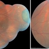
 Von Hippel Lindau with retinal capillary hemangioma
Nov 2 2023 by Marcelo Zas, MD PhD
30-year-old female patient diagnosed with Syndrome VHL (Von Hippel Lindau). Stage II. In the first wide-field retinography of the right eye we can observe the exophytic retinal hemangiomas, rounded, slightly delimited, located in the peripheral retina in the upper and lower temporal quadrants and due to the exudation produced by them, hard exudates are observed in the star hemisphere, affecting the macula.
Photographer: Mariano Cotic MD
Imaging device: Silverstone SS OCT Optos
Condition/keywords: abnormal retinal vessel
-
 Advanced Coats' Disease with Neovascular Glaucoma
Aug 21 2019 by Victor M Villegas, MD
Advanced Coats' Disease with neovascular glaucoma.
Photographer: Giselle Deoliveira, Bascom Palmer Eye Institute, University of Miami
Imaging device: RetCam III
Condition/keywords: abnormal retinal vessel, bullous retinal detachment, Coats' disease, diffuse lipid exudate, foveal hard exudates, neovascular glaucoma, pediatric retina
-
 AVM
AVM
Aug 21 2013 by Howard Schatz, MD
Twenty year old white female, 20/25 OU.
Condition/keywords: abnormal retinal vessel
-
 AVM
AVM
Aug 21 2013 by Howard Schatz, MD
Twenty five year old white female, right eye 20/20, left eye 20/80.
Condition/keywords: abnormal retinal vessel
-
 AVM
AVM
-
 AVM
AVM
-
 AVM
AVM
Aug 21 2013 by Howard Schatz, MD
Twenty three year old white male, 20/20 OU.
Condition/keywords: abnormal retinal vessel
-
 AVM
AVM
Aug 21 2013 by Howard Schatz, MD
Thirty eight year old female, right eye 20/20, left eye 20/200.
Condition/keywords: abnormal retinal vessel
-
 AVM
AVM
-
 AVM
AVM
-
 AVM
AVM
Aug 21 2013 by Howard Schatz, MD
Twenty seven year old white male, right eye 20/20, left eye 20/20.
Condition/keywords: abnormal retinal vessel
-
 AVM
AVM
Aug 21 2013 by Howard Schatz, MD
Fifty eight year old white female.
Condition/keywords: abnormal retinal vessel
-
 AVM
AVM
Aug 21 2013 by Howard Schatz, MD
Not an AV shunt, Idiopathic RV, superior nasal grads.
Condition/keywords: abnormal retinal vessel
-
 AVM
AVM
Aug 21 2013 by Howard Schatz, MD
Sixty year old white female, right eye 20/20, left eye 10/200, venous loop.
Condition/keywords: abnormal retinal vessel
-
 AVM
AVM
-
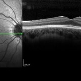
 Central Retinal Artery Occlusion with Cilioretinal Sparing - Optical Coherence Tomography
Oct 28 2020 by Fang Helen Mi
Optical coherence tomography showed hyper-reflective inner retinal layers, indicating intracellular oedema of the affected retina, with normal retinal layers in the area perfused by the cilioretinal artery.
Condition/keywords: central retinal artery occlusion (CRAO), cilioretinal sparing
-
 CHRPE
CHRPE
Jan 15 2021 by Priya Rasipuram Chandrasekaran, MBBS, DO, DNB, FRCS
This is the fundus photo and fundus photo montage of the left eye of a 25-year-old male showing flat, solitary, round, greyish pigmented lesion situated AT THE equator with a scalloped margin. Vessels overlying the lesion are normal and there is a clear demarcation line between this and normal retina. The margins are hypopigmented with few hypopigmented lacunae inside.
Condition/keywords: congenital hypertrophy of the retinal pigment epithelium (CHRPE)
-
 Combined Hamartoma of Retina and RPE
Jul 15 2020 by Itzel Ocampo
Fundus photograph of a 53-year-old man with a combined hamartoma of retina and RPE; visual capacity of hand movement. No systemic associations.
Photographer: Itzel Ocampo, Universidad Autonoma de Mexico, Hospital General de Mexico "Eduardo Liceaga"
Condition/keywords: abnormal retina, combined hamartoma
-
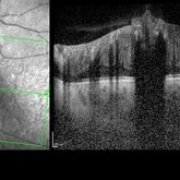 Combined Hamartoma of the Retina and Retinal Pigment Epithelium (CHRRPE)
Jan 21 2020 by Pierre-Henry Gabrielle, MD
Coupled OCT B-scan and IR imaging of a 17-year-old man with Combined hamartomas of the retina and retinal pigment epithelium (CHRRPE) at the posterior pole of the left eye. One can see a highly reflective elevated macular lesion with hyporeflective shadowing of the underlying tissue and obscuration of the normal retinal layers.
Photographer: Pierre-Henry Gabrielle, Ophthalmology department, Dijon University Hospital, France
Imaging device: Heidelberg Spectralis
Condition/keywords: combined hamartoma, optical coherence tomography (OCT)
-
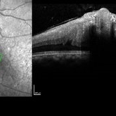 Combined Hamartoma of the Retina and Retinal Pigment Epithelium (CHRRPE)
Jan 21 2020 by Pierre-Henry Gabrielle, MD
Coupled OCT B-scan and IR imaging of a 17-year-old man with combined hamartomas of the retina and retinal pigment epithelium (CHRRPE) at the posterior pole of the left eye. One can see a highly reflective elevated macular lesion with hyporeflective shadowing of the underlying tissue and obscuration of the normal retinal layers.
Photographer: Pierre-Henry Gabrielle, Ophthalmology department, Dijon University Hospital, France
Imaging device: Heidelberg Spectralis
Condition/keywords: combined hamartoma, optical coherence tomography (OCT)
-
 Congenital Retinal Vessel Tortuosity
Apr 2 2024 by Pablo Angel Garcia Uribe
Fundus photograph of a 29-year-old man with bilateral congenital retinal vessel tortuosity. This image shows the sinuous course of retinal arterioles and a shiny internal limiting membrane.
Photographer: Pablo Ángel García-Uribe, Clínica Oftalmológica Salauno, Mexico City
Imaging device: NIDEK OCT RS-330 Duo 2
Condition/keywords: abnormal retinal vessel, anomalous vessels, Retina, tortuous vessels
-
 CSNB-OCT-OD
Aug 23 2021 by Jennifer Carstens
OCT/infrared image showing myopic fundus with normal retinal structure in patient with CACNA1F associated X-linked CSNB (OD).
Photographer: Jing Zhang, Ophthalmic Photographer
Condition/keywords: congenital stationary night blindness (CSNB), infrared image, optical coherence tomography (OCT)
 Loading…