Search results (153 results)
-
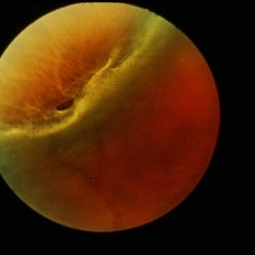
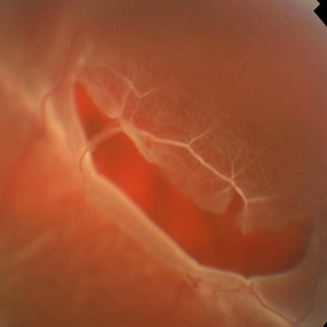 Retinal Tear at the Posterior Edge of Lattice Degeneration
Mar 1 2014 by Homayoun Tabandeh, MD, FASRS
Retinal tear at the posterior edge of lattice degeneration.
Condition/keywords: lattice degeneration, retinal tear
-
 Flat Lattice Lesion
Nov 9 2012 by Norman Byer
This 24-year-old woman had a flat lattice lesion without holes observed with no change for six years. She then developed two tiny retinal holes in this lesion and three years later the clinical retinal detachment shown here. She responded well to surgery. Even though such atrophic holes and lattice lesions may occasionally lead to a clinical detachment, it is important to understand that the mere presence of such holes is not an indication for prophylactic treatment. The reason for this is that we now know statistically that fewer than 1 percent of such cases lead to a retinal detachment.
Condition/keywords: lattice degeneration, retinal hole, scleral depression
-
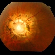
 Laser Treated Lattice Degeneration
Jul 12 2021 by Gabriel Costa Andrade, PhD
Fundus photograph of an 22-year-old man with peripheral lattice retinal degeneration treated with photocoagulation.
Photographer: Gabriel Andrade
Condition/keywords: lattice degeneration, retina
-
---thumb.JPG/image-square;max$300,300.ImageHandler) Lattice Degeneration
Lattice Degeneration
Jul 12 2013 by Jason S. Calhoun
Composite of HD-OCT and fundus photo showing demarcation line of lattice degeneration inferiorly temporally at 4-o'clock in a young black female.
Photographer: Jason S. Calhoun, Department of Ophthalmology, Mayo Clinic Jacksonville, Florida
Condition/keywords: lattice degeneration
-
 Lattice Degeneration
May 2 2013 by Henry J. Kaplan, MD
Pigmented lattice degeneration with lattice "wicker" caused by sclerotic blood vessels.
Condition/keywords: lattice degeneration, peripheral retinal degeneration
-
---thumb.jpg/image-square;max$300,300.ImageHandler) Lattice Degeneration
Lattice Degeneration
-
 Lattice Degeneration
Jan 5 2015 by H. Michael Lambert, MD
Lattice degeneration (stereo pair B).
Condition/keywords: lattice degeneration
-
 Lattice Degeneration
Jan 5 2015 by H. Michael Lambert, MD
Retinal detachment with lattice degeneration and holes (stereo pair A).
Condition/keywords: lattice degeneration
-
 Lattice Lesion
Nov 9 2012 by Norman Byer
This lattice lesion in a 36-year-old woman has remained unchanged over a period of 13 years. It shows a moderate snailtrack feature with discrete yellow dots visible on the surface of the lesion and especially along the posterior border. One of these can be well seen just below the lesion superimposed over the dark shadow of the scleral indentation. The exact nature of these yellow dots is still not entirely clear.
Condition/keywords: lattice degeneration, moderate snail track, scleral indentation, yellow dots
-
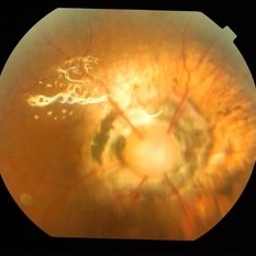
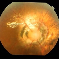
Peripheral Retinal Hole with OCT Co-localization
Sep 26 2023 by Bradley T. Smith, MD, FASRS
Peripheral asymptomatic atrophic retinal hole with OCT co localization demonstrating small cuff of sub retinal fluid. Near infrared imaging shows hyper reflectivity through hole.
Condition/keywords: atrophic hole, lattice degeneration, OCT
-
 Retinal Detachment and Lattice Degeneration
Mar 25 2025 by Korey Starkey
26 year-old patient presented at first visit with rhegmatogenous macula involving retinal detachment of the left eye. Underwent prompt surgical repair. Both eyes also present with lattice degeneration with atrophic holes.
Photographer: Korey Starkey
Condition/keywords: atrophic retinal hole, fundus photography, lattice degeneration, montage photo, Optos, OPTOS CALIFORNIA RGB, retinal detachment, retinal holes, rhegmatogenous retinal detachment, ultra-wide field imaging
-
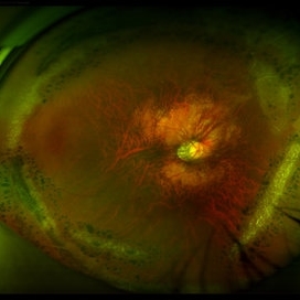
 Morning Glory Disc
Apr 22 2016 by Mallika Goyal, MD
Right fundus of a 34-year-old lady with bilateral morning glory disc anomaly with silicon oil in-situ; this eye had rhegmatogenous retinal detachment with multiple peripheral lattice degeneration and was successfully operated. However, there was redetachment within a week of silicon oil removal in absence of any untreated retinal breaks suggesting the abnormal disc as a likely cause of the redetachment.
Photographer: Mallika Goyal, MD, Apollo Health City, Hyderabad, India
Condition/keywords: Morning Glory Syndrome
-
 Lattice Degeneration and Choroidal Nevus
Oct 10 2015 by Hamid Ahmadieh, MD
Color fundus photograph of the right eye of a 46-year-old woman with a typical lattice degeneration and an adjacent choroidal nevus.
Photographer: Solmaz Shahmohammad, Negah Eye Center, Tehran, Iran
Condition/keywords: choroidal nevus, color fundus photograph, lattice degeneration
-
 Lattice Lesion
Nov 9 2012 by Norman Byer
In this 47-year-old woman, this lattice lesion with a small hole in the right end has led to a subclinical retinal detachment which extends to the margin of the subtle yellowish zone almost at the upper edge of the photograph. This patient did not desire surgery, and the area of detachment has changed only a small amount in the past eight years. The risk of a clinical retinal detachment developing from lattice degeneration is less than 1 percent. In those cases where it does though, about 3 quarters are caused by a tractional tear and about one quarter are caused by an atrophic hole as in this case.
Condition/keywords: atrophic retinal hole, lattice degeneration, lattice lesion, retinal hole, yellowish zone
-
 Morning Glory Disc
Apr 22 2016 by Mallika Goyal, MD
Right fundus of a 34-year-old lady with bilateral morning glory disc anomaly with silicon oil in-situ; this eye had rhegmatogenous retinal detachment with multiple peripheral lattice degeneration and was successfully operated. However, there was redetachment within a week of silicon oil removal in absence of any untreated retinal breaks suggesting the abnormal disc as a likely cause of the redetachment.
Photographer: Mallika Goyal, MD, Apollo Health City, Hyderabad, India
Condition/keywords: Morning Glory Syndrome
-
 Retinal Break at Site of Lattice Degeneration with Scleral Indentation
Nov 9 2012 by Norman Byer
This is the same case as the previous photograph. With scleral indentation slightly more posterior, the flap is seen to be associated with a large retinal tear. This is a tractional tear and it is possible that in this case the cryotherapy itself may have increased the vitreoretinal traction at this site and in this way led to this new tear. The age of the tear is unknown because it was asymptomatic, and even though the eye is aphakic the tear has not caused a clinical retinal detachment.
Condition/keywords: retinal flap, scleral indentation, tractional retinal tear, vitreoretinal traction
-
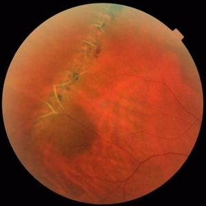
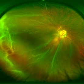 Rhegmatogeous Retinal Detachment
Mar 21 2013 by Yusuke Oshima, MD, PhD
Wide-field fundus photograph of a 38-year-old woman with a macula-involved retinal detachment due to a tiny break localized around the edge of a lattice degeneration.
Photographer: Yusuke Takada, Osaka University Graduate School of Medicine
Imaging device: OPTOS 200Tx
-
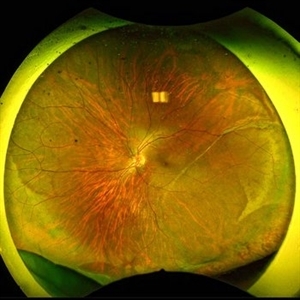
 Laser Barrage for Temporal Localized Rhematogenous Retinal Detachment
Feb 15 2018 by Kushal S Delhiwala, MBBS, MS, FMRF,FICO, FAICO
39-year-old female presenting with sudden onset flashes and floaters in left eye having undergone refractive surgery 20 years before for pathologic myopia.Color fundus photograph montage of left eye showing macula sparing inferotemporal localized Rhematogenous retinal detachment with horse shoe tear and temporal lattice degeneration treated with laser barrage.
Photographer: Dr Kushal Delhiwala, Netralaya superspeciality eye hospital ,Ahmedabad
Imaging device: Zeiss Visucam 500
Condition/keywords: barrier laser, macula sparring
-
 Macula-Sparing GRT RRD
Jul 6 2017 by Andrew A. Moshfeghi, MD, MBA, FASRS
Wide-field fundus photograph of a 43-year-old myopic man with a history of lattice retinal degeneration status posterior barrier laser performed elsewhere who presented with a giant-retinal tear associated retinal detachment of the right eye.
Photographer: Jay Jiang, University of Southern California Roski Eye Institute
Imaging device: Optos California
Condition/keywords: acute retinal detachment, giant retinal tear, lattice degeneration
-
 Morning Glory Disc
Apr 22 2016 by Mallika Goyal, MD
Left fundus of a 34-year-old lady with bilateral morning glory disc anomaly; fellow eye had rhegmatogenous retinal detachment with multiple peripheral lattice degeneration and was successfully operated.
Photographer: Mallika Goyal, MD, Apollo Health City, Hyderabad, India
Condition/keywords: Morning Glory Syndrome
-
 1 year Follow Up after Scleral Buckle Surgery in a Young Patient
May 18 2023 by Jesus Lozano, MD
25 year old man after Scleral Buckle Surgery + laser Retinopexy do to RRD macula off with ínfero temporal mid peripheral retinal holes in an area of lattice degeneration. Final VA 6/9.
Imaging device: Optos
Condition/keywords: scleral buckle
-
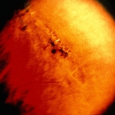
 Acute Posterior Vitreous Detachment
Nov 9 2012 by Norman Byer
This large and complicated retinal tear in a 51-year-old man resulted from an acute posterior vitreous detachment which concentrated its tractional forces around this area of lattice degeneration. Because of the powerful traction, there is an additional central tear splitting the large retinal flap and almost severing one of its arms. The traction was strong enough to completely rupture the blood vessel just to the left of the flap. Marking the ruptured peripheral end of the blood vessel is a yellow depigmented thrombus.
Condition/keywords: acute posterior vitreous detachment, depigmented thrombus, lattice degeneration, retinal tear, tractional retinal detachment
-
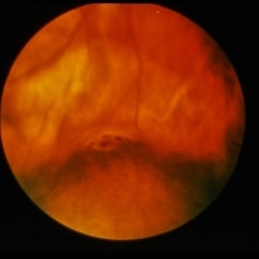
 Acute Retinal Detachment
Nov 9 2012 by Norman Byer
This 54-year-old man was referred because of sudden symptoms in his opposite eye in which he had suffered an acute retinal detachment secondary to a horseshoe tear around lattice degeneration. During the examination, the fellow eye shown here was also found to have this large horseshoe tear about 1 o’clock hour (4 disc diameters) in size. A tear occurred around a lattice lesion which is present on the flap but is out of focus. This tear had been asymptomatic even though it was caused by a posterior vitreous detachment and illustrates that even very large tears may produce no symptoms or mild symptoms that are easily overlooked.
Condition/keywords: lattice degeneration, posterior vitreous detachment
-
 Chronic Retinal Detachment with Proliferative Vitreoretinopathy
Jan 25 2024 by Isaac Agranoff
Widefield fundus photography of a 24 year old male presenting with subtotal retinal detachment with circumferential anterior proliferative vitreoretinopathy. The detachment is bullous inferiorly with atrophic retina and subretinal bands. There are also scattered patches of lattice with atrophic holes and associated detachment in the periphery. Patient presented with flashes for 2 years with worsening vision over the past 6-8 months, measured at 20/100 ph 20/60 OS.
Photographer: Isaac Agranoff, Ashley Rigdon
Imaging device: Optos California
Condition/keywords: atrophic hole, chronic retinal detachment, lattice degeneration, proliferative vitreoretinopathy (PVR), subretinal bands
-
 ERMageddon - Wrinkle in the Space-time Fabric of Macula
Oct 29 2025 by SHRADDHA RAJ SHRIVASTAVA
38 year old female with Epiretinal Membrane (ERM) over macula, post laser barrage for multiple symptomatic Horse-shoe Tears (HSTs) and Lattice Degenerations. Posterior pole revealed tilted disc with peripapillary atrophy. There is thick opaque epiretinal membrane obscuring the underlying superior arcade vessels and causing foveal ectopia with distortion of perimacular vasculature. Patient was planned for Right Eye pars plana vitrectomy for ERM peeling.
Photographer: Dr. Shraddha Raj Shrivastava
Imaging device: Nidek Mirante SLO/OCT (Confocal scanning/Spectral domain OCT
Condition/keywords: ectopic fovea, epiretinal membrane (ERM), ERM, horseshoe tear, vitreomacular traction (VMT)
 Loading…