Search results (62 results)
-
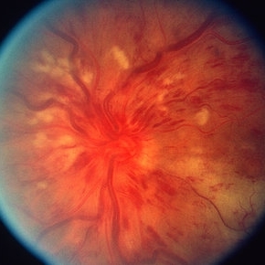 CRVO
CRVO
Mar 29 2013 by Henry J. Kaplan, MD
Full blown ischemic CRVO with disc swelling, dilated and tortous veins, scattered hemorrhages and multiple cotton wool spots.
Condition/keywords: central retinal vein occlusion (CRVO), ischemic CRVO
-
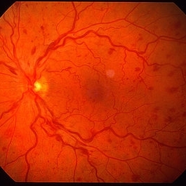 Non Ischemic CRVO
Mar 29 2013 by Henry J. Kaplan, MD
Non ischemic CRVO.
Condition/keywords: central retinal vein occlusion (CRVO)
-
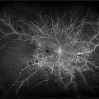
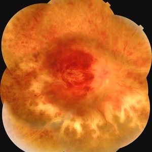
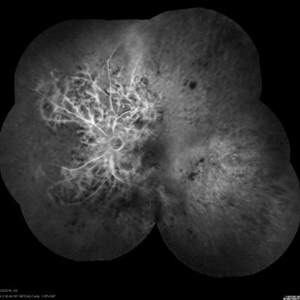
 Central Retinal Vein Occlusion with Retinal Neovascularization
Jan 19 2022 by Olivia Rainey
Ultra-widefield fluorescein angiogram of a 56-year-old male with a Central Retinal Vein Occlusion with Retinal Neovascularization affecting his left eye. The patient presented on 1/19/2022 with scNLP vision in the left eye. The patient has good PRP, however areas of ischemia still remain untreated by laser. He also has severe neovascular glaucoma contributing to his poor vision.
Photographer: Olivia Rainey, OCT-C, COA
Imaging device: Optos California
Condition/keywords: central retinal vein occlusion (CRVO), FA early phase, fluorescein angiogram (FA), hemorrhage, ischemic CRVO, left eye, neovascular glaucoma, Optos, pan-retinal photocoagulation (PRP), retinal ischemia, retinal neovascularization, ultra-wide field imaging
-
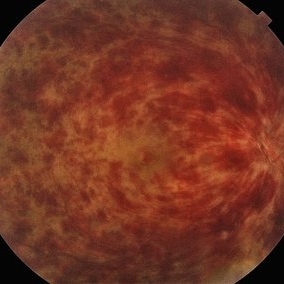
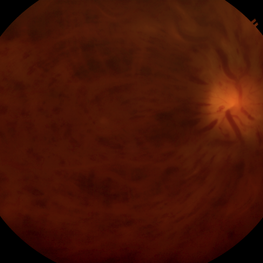
 Central Retinal Vein Occlusion with Severe Retinal Ischemia
Jan 19 2022 by Olivia Rainey
Ultra-widefield fluorescein angiogram of a 56-year-old male with a Central Retinal Vein Occlusion with Severe Retinal Ischemia affecting his right eye. The patient presented on 1/19/2022, sc20/20-2 vision in the right eye. The patient has had a good response to Eylea with complete resolution of edema. The physician is considering PRP to ischemic periphery in the future and given the degree of ischemia in both eyes, she recommends that the patient's PCP check carotid Doppler US.
Photographer: Olivia Rainey, OCT-C, COA
Imaging device: Optos California
Condition/keywords: central retinal vein occlusion (CRVO), FA late phase, fluorescein angiogram (FA), ischemic CRVO, Optos, retinal ischemia, ultra-wide field imaging
-
 CRVO with Secondary CLRAO
May 28 2020 by Richard M Martindale, MD
Non-ischemic CRVO (VA 20/30) with secondary CLRAO (nasal macular pallor) in a hypertensive 69yo female. Pathophysiologically, the cilioretinal artery occlusion occurs secondary to elevation in the hydrostatic pressure in the retinal venous system relative to the choroidal perfusion pressure (which supplies the cilioretinal artery).
Photographer: Retina Consultants of Alabama, P. C.
Imaging device: Optos
Condition/keywords: cilioretinal artery occlusion, non-ischemic central retinal vein occlusion (CRVO)
-
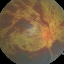
 Ischemic Central Retinal Vein Occlusion
Aug 6 2024 by César Adrián Gómez Valdivia, MD
Fundus photograph of an 80 year old man with and acute central retinal vein occlusion, ischemic variant.
Photographer: @eyemissu2
Condition/keywords: central retinal vein occlusion (CRVO), ischemic CRVO
-
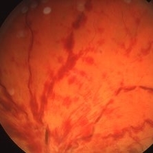
 OLD ISCHEMIC CRVO
Jun 11 2022 by Nivesh Gupta
Central fundus image showing tortuous vessels.
Photographer: DR. NIVESH GUPTA, RETINA FELLOW , RETINA FOUNDATION, AHMEDABAD
Imaging device: NIDEK MIRANTE
Condition/keywords: ischemic CRVO
-
 Ischemic Central Retinal Vein Occlusion
Jan 24 2019 by Nichole Lewis
76-year-old woman with an ischemic central retinal vein occlusion, severely attenuated and sclerotic vessels and scattered retinal hemorrhages. Vision decrease over 1 year. VA 20/CF. Patient is returning for pan retinal photocoagulation.
Photographer: Nichole Lewis
Imaging device: Optos
Condition/keywords: attenuated vessels, central retinal vein occlusion (CRVO), hemorrhage, ischemic CRVO, sclerotic vessels
-
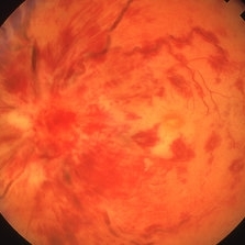
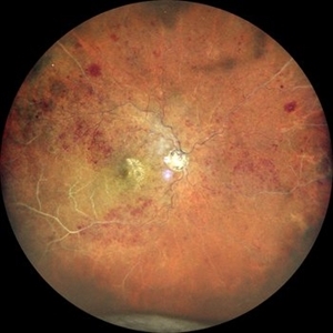
 A Vein in Vain: Ischemic CRVO
Dec 6 2024 by Jasmeet Kaur Chandi
Fundus photo of a 55 year-old female with extensive flame-shaped and dot-blot hemorrhages in all four quadrants. Tortuous and dilated veins with cotton-wool spots. Optic disc swelling with hyperemia and macular edema.
Photographer: Dr. Jasmeet Kaur Chandi
Condition/keywords: Ischemic Central Retinal Vein Occlusion
-
 AIDS and CRVO
Mar 26 2019 by Gary R. Cook, MD, FACS
43-year-old patient with AIDS and non-perfused CRVO; Visual Acuity = NLP.
Imaging device: Topcon VT-50
Condition/keywords: AIDS, ischemic CRVO
-
 AIDS and CRVO
Mar 26 2019 by Gary R. Cook, MD, FACS
43-year-old patient with AIDS and a non-perfused CRVO; Visual Acuity = NLP.
Imaging device: Topcon VT-50
Condition/keywords: AIDS, ischemic CRVO
-
 Angle Neovascularization
Mar 21 2013 by Yusuke Oshima, MD, PhD
Angle neovascularization due to ischemic CRVO.
Photographer: Yusuke Oshima, MD, PhD, Osaka University Graduate School of Medicine
Condition/keywords: angle neovascularization, gonioscopy
-
 Central Retinal Vein Occlusion
Dec 22 2018 by FELIPE PEREIRA
25-year-old male patient with acute and painless vision loss of left eye. The fundus examination demonstrate optic disc swelling, venous tortuosity, diffuse intraretinal hemorrhage and severe macular edema. There is also extensive exudative retinal detachment with lipid deposits in the posterior pole, mainly around the vessels. The systemic work up was negative, including serologies, rheumatologic and hematological markers and cholesterol and triglycerides within normal limits.
Photographer: Felipe Pereira
Imaging device: Vizucan, Zeiss
Condition/keywords: central vein occlusion, ischemic CRVO
-
 Central Retinal Vein Occlusion
Jan 28 2024 by Gayathri Mohan
Fundus photograph of a 50 year old woman showing a CRVO.
Photographer: Dr Gayathri Mohan, Thumbay Medical and Dental Speciality Centre, U.A.E
Imaging device: 3nethra
Condition/keywords: central retinal vein occlusion (CRVO), ischemic CRVO
-
 Central Retinal Vein Occlusion
Apr 9 2024 by Akansha Sharma
Color fundus photograph of a 73 year old hypertensive male with central retinal vein occlusion.
Photographer: Dr. Akansha Sharma, Bharati Eye Hospital
Condition/keywords: central retinal vein occlusion (CRVO), ischemic CRVO
-
 Central Retinal Vein Occlusion
Apr 9 2024 by Akansha Sharma
Color fundus photograph of a 73 year old hypertensive male with central retinal vein occlusion.
Photographer: Dr. Akansha Sharma, Bharati Eye Hospital
Condition/keywords: central retinal vein occlusion (CRVO), ischemic CRVO
-
 Central Retinal Vein Occlusion (CRVO)
Jun 19 2020 by Stephanie Burke
85-year-old male with ischemic CRVO.
Photographer: Stephanie Burke, CRA, OCT-C
Condition/keywords: central retinal vein occlusion (CRVO), ischemia, macular edema, ultra-wide field imaging
-
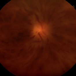
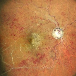
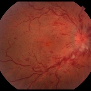 Central Retinal Vein Occlusion (CRVO) Associated with Papillophlebitis
Apr 16 2021 by Gabriel Costa Andrade, PhD
Fundus photograph of an 38-year-old woman with Central retinal vein occlusion (CRVO) associated with papillophlebitis.
Photographer: Dr Gabriel Andrade
Condition/keywords: central retinal vein occlusion (CRVO), ischemic CRVO
-
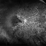
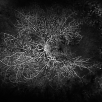
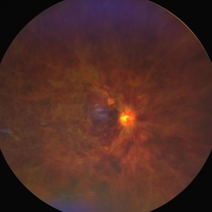
 Central Retinal Vein Occlusion with Severe Ischemia
May 22 2016 by Olivia Rainey
Composite fluorescein angiogram of the left eye of a male with a Central Retinal Vein Occlusion with severe ischemia.
Photographer: Olivia Rainey
Imaging device: Heidelberg Spectralis
Condition/keywords: central retinal vein occlusion (CRVO), composite, fluorescein leakage, ischemic CRVO
-
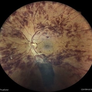
 Central vein occlusion (chronic)
Nov 26 2023 by Anjana Mirajkar, MS Ophthalmology
A widefield color photo image of RE of a 60 year old male in a case of central vein occlusion.
Photographer: Dr. Anjana Mirajkar -Retina Foundation, Ahmedabad
Imaging device: Mirante-Nidek
Condition/keywords: central retinal vein occlusion (CRVO), central vein occlusion, ischemic CRVO
-
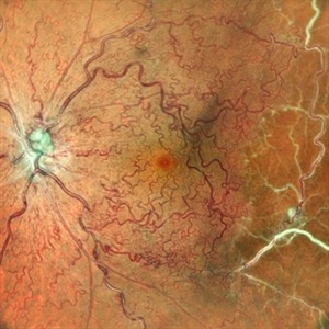
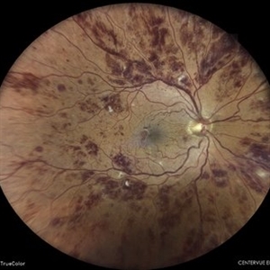
 Central vein occlusion (chronic)
Nov 26 2023 by Anjana Mirajkar, MS Ophthalmology
A central color photo image of RE of a 60 year old male in a case of central vein occlusion.
Photographer: Dr. Anjana Mirajkar -Retina Foundation, Ahmedabad
Imaging device: Mirante-Nidek
Condition/keywords: central retinal vein occlusion (CRVO), ischemic CRVO
-
 Combined central retinal vein and artery occlusion
Jun 19 2022 by T. P . VIGNESH, MBBS,MS
Fundus photo of a 67 year old female patient, revealing multiple superficial retinal haemorrhages and occlusion of both central retinal vein and central retinal artery.
Photographer: Bharathi Singaravel
Imaging device: Zeiss Clarus
Condition/keywords: central retinal artery occlusion (CRAO), COMBINED, ischemic CRVO
-
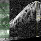 CRVO
CRVO
May 17 2024 by T. P . VIGNESH, MBBS,MS
SD- OCT of the right eye of a 57 year old man with Ischaemic CRVO and recurrent chronic CME .
Photographer: Sivanath
Imaging device: Heidelberg Spectralis
Condition/keywords: ischemic CRVO
-
 CRVO
CRVO
 Loading…