Search results (57 results)
-
 Silicon Oil
Apr 27 2018 by Giselle DeOliveira
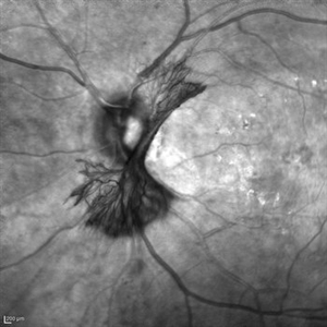
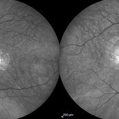
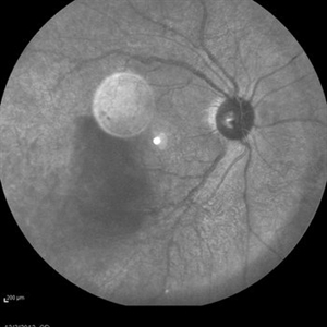
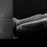
Infrared photo of 75-year-old male with retinal detachment.
Photographer: Giselle DeOliveira, University of Miami, Bascom Palmer Eye Institute
Imaging device: Heidelberg Spectralis
Condition/keywords: infrared image, silicone oil
-
 Acute Macular Neuroretinopathy
Dec 11 2019 by Lauren Whaley
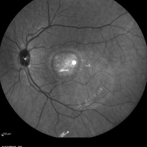
34-year-old female patient presented with changes in vision after recent upper respiratory infection. Referring doctor originally thought it was a blood pressure issue. She noticed a "C" shape in her vision. Infrared image was captured showing exactly what patient was describing! Doctor confirmed with this image that it was AMN.
Photographer: Lauren R. Whaley, COA
Imaging device: Heidelberg Spectralis
Condition/keywords: 30 degrees, acute macular neuroretinopathy, Heidelburg Spectralis, left eye, macula, near infrared autofluorescence (NIRAF)
-
 Cuticular and soft drusen
Jun 14 2021 by Gerardo Garcia-Aguirre, MD
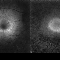
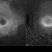
Fundus photograph (left) and Retro mode infrared image (right) of an eye with soft and cuticular drusen. Drusen are highlighted and better visualized with retro mode imaging.
Photographer: Gerardo Garcia-Aguirre
Imaging device: Nidek Mirante
Condition/keywords: drusen, dry age-related macular degeneration (dry AMD)
-
 "NVD Flower"
Oct 20 2023 by Daniel Davis, OCT-C
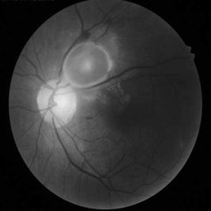
Infrared image of NVD (52F)
Imaging device: Heidelberg Spectralis
Condition/keywords: neovascularization of the disc (NVD)
-
 Best Disease
Mar 9 2013 by Hamid Ahmadieh, MD
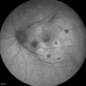
Infrared imaging of the left eye of a 49-year-old man with decreased VA due to advanced Best disease.
Photographer: Soodabeh Fooladin, Negah Eye Center, Tehran
Imaging device: Heidelberg Spectralis
Condition/keywords: Best disease, infrared image
-
 Case 2 Retinitis Pigmentosa BAF IRAF OD
May 14 2014 by Avris Romario Diparaja Siahaan
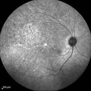
Fundus image a 57-year-old man with retinitis pigmentosa on both eyes. These image were taken with blue auto fluorescein mode (BAF) and infrared auto fluorescence (IRAF).
Photographer: Avris Romario Diparaja Siahaan
Imaging device: Heidelberg HRA + OCT Spectralis
Condition/keywords: autofluorescence imaging, fundus photograph, infrared image, retinitis pigmentosa
-
 Case 2 Retinitis Pigmentosa BAF IRAF OS
May 14 2014 by Avris Romario Diparaja Siahaan
Fundus image a 57-year-old man with retinitis pigmentosa on both eyes. These image were taken with blue auto fluorescein mode (BAF) and infrared auto fluorescence (IRAF).
Photographer: Avris Romario Diparaja Siahaan
Imaging device: Heidelberg HRA + OCT Spectralis
Condition/keywords: autofluorescence imaging, fundus photograph, infrared image, retinitis pigmentosa
-
 Central Areolar Choroidal Dystrophy
Jul 7 2015 by Hamid Ahmadieh, MD
Infrared image of both eyes of a 58-year-old man with progressive loss of vision. VA OD is 20/60 and VA OS is 20/400.
Photographer: Soulmaz Shahmohammad, Negah Eye Center, Tehran, Iran
Imaging device: Specteralis
Condition/keywords: central areolar choroidal dystrophy (CACD), infrared image
-
 Macular Tear
May 14 2014 by Avris Romario Diparaja Siahaan
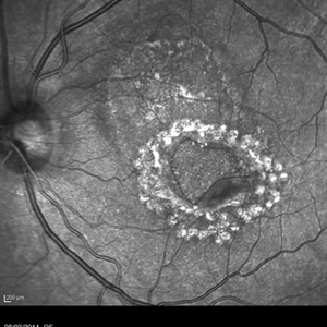
Infrared photograph a 40-year-old man with macular tear (had a photocoagulation laser).
Photographer: Avris Romario Diparaja Siahaan
Imaging device: Heidelberg HRA + OCT Spectralis
Condition/keywords: infrared image, macular hole
-
 Angioid Streaks With Associated Disc Drusen and CNV
Sep 21 2018 by Sarah Oelrich
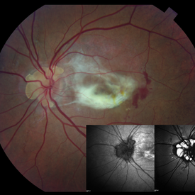
Angioid streaks with associated disc drusen and CNV.
Photographer: Sarah Oelrich CRA COT, Southeastern Retina Associates Knoxville Tn
Condition/keywords: angioid streaks, autofluorescence imaging, choroidal neovascularization (CNV), disc drusen, infrared image
-
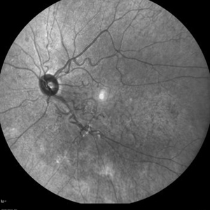
 Proliferative Diabetic Retinopathy
Sep 15 2012 by Hamid Ahmadieh, MD
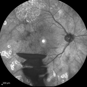
Infrared image of a 30-year-old woman with the history of scatter laser photocoagulation and a preretinal hemorrhage due to active PDR .
Photographer: Hamid Ahmadieh, MD, Ophthalmic Research Center, Labbafinejad Medical Center, Shahid Beheshti University of Medical Sciences
Imaging device: Heidelberg HRA
Condition/keywords: infrared image, preretinal hemorrhage
-
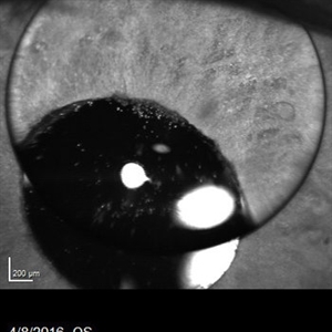 Silicone Oil Bubble in Anterior Chamber - 15 Degree Angle
Apr 11 2016 by Zach Dupureur
30 % silicone oil bubble involving central visual axis. Occurred after a PPV with silicone oil. Oil from vitreous moved into the anterior chamber.
Photographer: Zachary Dupureur, OCT-C
Imaging device: Heidelberg Spectralis
Condition/keywords: anterior chamber, detachment, infrared image, pars plana vitrectomy (PPV), scleral buckle, silicone oil
-
 Behcet's Disease
Mar 13 2013 by Hamid Ahmadieh, MD
Infrared image of the right eye of a 23-year-old man with retinal vasculitis and branch retinal vein occlusion (BRVO) due to Behcet's disease .
Photographer: Solmaz Shahmohammad, Negah Eye Center, Tehran
Imaging device: Heidelberg Spectralis
Condition/keywords: branch retinal vein occlusion (BRVO), infrared image, retinal vasculitis
-
 Central Retinal Vein Occlusion
Oct 7 2015 by Avris Romario Diparaja Siahaan
An IR + OCT image of a 46-year-old man with a central retinal vein occlusion on his left eye.
Photographer: Avris Romario Diparaja Siahaan, Klinik Mata Nusantara
Imaging device: Spectralis Heidelberg
Condition/keywords: central retinal vein occlusion (CRVO), infrared image, optical coherence tomography (OCT)
-
 Choroidal Melanoma
Feb 2 2018 by Olivia Rainey
Optical coherence tomography with enhanced depth imaging of a 78-year-old female with choroidal melanoma with subretinal fluid affecting her right eye.
Photographer: Olivia Rainey
Imaging device: Heidelberg Spectralis
Condition/keywords: enhanced depth imaging, infrared image, optical coherence tomography (OCT), subretinal fluid, superior retina
-
 Combined Hamartoma of the Retina and Retinal Pigment Epithelium (CHRRPE)
Jan 21 2020 by Pierre-Henry Gabrielle, MD
IR imaging of a 17-year-old man with Combined hamartomas of the retina and retinal pigment epithelium (CHRRPE) at the posterior pole of the left eye.
Photographer: Pierre-Henry Gabrielle, Ophthalmology department, Dijon University Hospital, France
Imaging device: Heidelberg Spectralis
Condition/keywords: combined hamartoma, infrared image
-
 Cone-Rod Dystrophy
Mar 15 2017 by Hamid Ahmadieh, MD
Infrared and OCT images of the left eye of a 16-year-old boy with decreased visual acuity and color vision deficiency due to cone-rod dystrophy.
Photographer: Abazarnezhad , Negah Eye Center, Tehran, Iran
Imaging device: Spectralis OCT
Condition/keywords: cone dystrophy, infrared image, optical coherence tomography (OCT)
-
 Cone-Rod Dystrophy
Mar 15 2017 by Hamid Ahmadieh, MD
Infrared and OCT images of the right eye of a 16-year-old boy with decreased visual acuity and color vision deficiency due to cone-rod dystrophy.
Photographer: Abazarnezhad , Negah Eye Center, Tehran, Iran
Imaging device: Spectralis OCT
Condition/keywords: cone dystrophy, infrared image, optical coherence tomography (OCT)
-
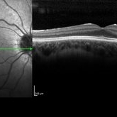
CSNB-OCT-OD
Aug 17 2021 by Christine Kay, MD
This is an OCT/infrared image OD exhibiting normal fundus in a 16 year-old male with X-linked CSNB with proven mutation in CACNA1F.
Photographer: Christine Kay, MD
Condition/keywords: infrared image, X-linked CSNB
-
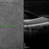
 CSNB-OCT-OD
Aug 23 2021 by Jennifer Carstens
OCT/infrared image showing myopic fundus with normal retinal structure in patient with CACNA1F associated X-linked CSNB (OD).
Photographer: Jing Zhang, Ophthalmic Photographer
Condition/keywords: congenital stationary night blindness (CSNB), infrared image, optical coherence tomography (OCT)
-
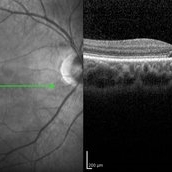
 CSNB-OCT-OS
Aug 23 2021 by Jennifer Carstens
OCT/infrared image showing myopic fundus with normal retinal structure in patient with CACNA1F associated X-linked CSNB (OS).
Photographer: Jing Zhang, Ophthalmic Photographer
Condition/keywords: congenital stationary night blindness (CSNB), infrared image, optical coherence tomography (OCT)
-
CSNB-OCT-OS
Aug 17 2021 by Christine Kay, MD
This is an OCT/infrared image of the left eye exhibiting normal fundus in a 16 year-old male with X-linked CSNB with proven mutation in CACNA1F.
Photographer: Christine Kay, MD
Condition/keywords: X-linked CSNB
-
 Cysticercosis Cyst
Apr 29 2019 by Chintan Sarvaiya, MS
Infrared image of a 43-year-old male with Cysticercosis cyst
Photographer: Dr. Chintan Sarvaiya, Banker's Retina Clinic
Condition/keywords: cysticercosis
-
 Endogenous Endophthalmitis
Sep 3 2014 by Hamid Ahmadieh, MD
Infrared image of the left eye of a 45-year-old diabetic man with the history of urinary tract infection. The most probable diagnosis was candida endogenous endophthalmitis.
Photographer: Nayereh Hadipour, Negah Eye Center, Tehran, Iran
Condition/keywords: candida endophthalmitis, endogenous endophthalmitis, infrared image
-
 Fundus Flavimaculatus and CNV
Nov 14 2013 by Hamid Ahmadieh, MD
Infrared image of the right eye of a 35-year-old woman with subfoveal CNV secondary to fundus flavimaculatus .
Photographer: Nayereh Hadipour, Negah Eye Center, Tehran
Condition/keywords: choroidal neovascularization (CNV), fundus flavimaculatus, infrared image, retinal flecks
 Loading…