Search results (131 results)
-
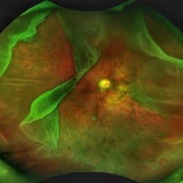
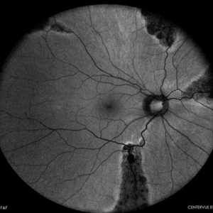
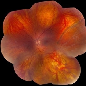
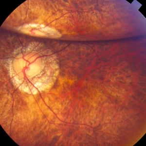 "Internal Mirroring" Effect by Intraocular Gas
Mar 25 2014 by Homayoun Tabandeh, MD, FASRS
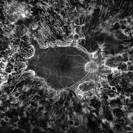
"Internal mirroring" by residual intraocular gas in a highly myopic patient 3 weeks post repair of retinal detachment with pars plana vitrectomy and C3F8 gas.
Photographer: Danny Rivas
Condition/keywords: high myopia, intraocular gas
-
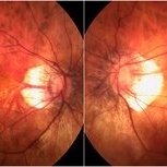
 Foveoschisis secondary to high myopia
Mar 13 2015 by Niloofar Piri, MD
Infrared and HD-OCT of the right eye in a 55-year-old African American female with high myopia (more than -6.00 D), BCVA: 20/25 OU Cartwheel appearance of the fovea in the infrared imaging is visible. HD- OCT demonstartes schisis in different layers of the retina (both NFL and OPL; notice stretching of the Muller cells); VMT is also present . Outer retinal layers are preserved which explains the good vision . She had the same findings in OS.
Photographer: Niloofar Piri, MD
Imaging device: Heidelberg Spectralis
Condition/keywords: high myopia, retinoschisis
-
 Myelinated Nerve Fibre (MNF)
Jun 17 2023 by Harsh Vardhan Singh, MS
Fundus photograph of 32-year-old male having good best corrected visual acuity in both eyes with right eye having high myopia & MNF as incidental finding
Photographer: Dr Harsh Vardhan Singh, Assistant Professor, AIIMS, Guwahati
Condition/keywords: medullated nerve fibers, MNF, myelinated nerve fiber layer, myelinated nerve fibers, Nerve fiber layer arrangements, NFL
-
 Prominent Long Ciliary Nerve
Jan 25 2022 by Kachelle Brown
Ultra-wide field photograph of a 48-year-old female with a prominent long ciliary nerve. Patient presented asymptomatic, and was referred for a macula on retinal detachment. Patient was diagnosed with high myopia and a posterior vitreous detachment, and the physician discussed increased risk of floaters, myopic degeneration and retinal detachment associated with high myopia. -24.00 prior to cataract surgery OU per patient.
Photographer: Kachelle Brown
Imaging device: Optos California
Condition/keywords: fundus photograph, high myopia, long ciliary nerve, optos, right eye, ultra-widefield image
-
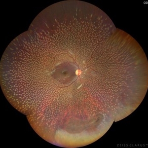 Benign Familial Fleck Retina
Dec 21 2023 by Vishal Agrawal, MD, FRCS,FACS,FASRS
10-year male with high myopia on examination revealed diffuse flecks distributed all over fundus in both eyes sparing macula. Inferior lattice with WWOP areas were also noted in right eye.
Photographer: Dr Ayushi
Imaging device: Clarus 700
Condition/keywords: fleck retinopathy, myopia
-
 Dome-Shaped Macula With Subretinal Fluid
Jun 14 2018 by Gerardo Garcia-Aguirre, MD
EDI OCT of the right eye of a 17-year-old highly myopic girl. Subfoveal fluid is present. There is choroidal thinning, and scleral thickening in the foveal area.
Photographer: Gerardo Garcia-Aguirre, MD
Imaging device: Heidelberg Spectralis
Condition/keywords: dome shaped macula, high myopia
-
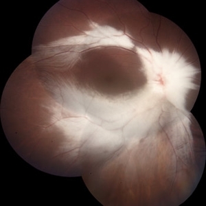
 Giant Retinal Tear with Multiple Retinal Breaks
Apr 21 2025 by Hrishikesh Naik, MS
A 28 year old high myope with retinal detachment associated with a supero-temporal giant retinal tear in addition to multiple peripheral horseshoe tears and an additional supero-nasal retinal tear.
Photographer: Hrishikesh Naik
Imaging device: Optos Daytona
Condition/keywords: giant retinal tear, High Myopia, horseshoe tear, retinal break, retinal detachment
-
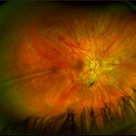
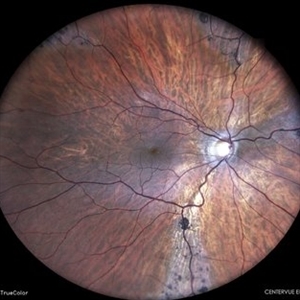
 High Myopia
Dec 7 2019 by Anfisa Ayalon, MD
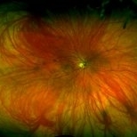
Fundus photograph of a 55-year-old woman with high myopia.
Photographer: Anfisa Ayalon,MD., Meir Medical Center, Kfar Saba, Israel.
Condition/keywords: high myopia, myopia, peripapillary atrophy
-
 Myelinated Nerve Fibers
Apr 18 2025 by DR Rohit Gupta
The **myelinated nerve fibers of the optic disc** (also known as **medullated nerve fibers**) are retinal nerve fibers that retain their myelin sheath as they pass through the optic nerve head. Normally, retinal nerve fibers are unmyelinated to allow for light transparency, but in some cases, myelination extends anteriorly into the retina, appearing as a striking white, feathery patch on the optic disc or peripapillary retina. ### **Key Features:** 1. **Appearance:** - Dense, white, striated patches with feathery edges. - Typically located at the superior or inferior pole of the optic disc. - May obscure retinal vessels underneath. 2. **Clinical Significance:** - Usually **benign** and asymptomatic. - **Congenital** (present at birth or early childhood). - Rarely associated with **visual field defects** (e.g., scotomas corresponding to the area of myelination). - Occasionally linked with **high myopia** or **amblyopia** if extensive. 3. **Pathophysiology:** - Failure of oligodendrocytes or Schwann cells to stop myelination at the lamina cribrosa. - Normally, myelination stops at the optic nerve head, but in this condition, it extends into the retina. 4. **Diagnosis:** - **Fundoscopy:** Classic white, feathery appearance. - **Optical Coherence Tomography (OCT):** Shows thickened retinal nerve fiber layer (RNFL). - **Visual Field Testing:** May detect defects if large. 5. **Differential Diagnosis:** - Optic disc edema - Cotton wool spots - Retinoblastoma (rarely, but must be ruled out in children) 6. **Management:** - No treatment required if asymptomatic. - Monitor for amblyopia in children. - Rare cases with significant visual impairment may need further evaluation. ### **Fun Fact:** Myelinated nerve fibers are seen in **~0.5-1%** of the population and are usually an incidental finding.
Photographer: Dr Rohit gupta
Imaging device: Samsung S21
Condition/keywords: Medulated Nerve fibre, Medullated Nerve fibres, myelinated nerve fibers, Myelinated Nerve Fibres, optic disc drusen
-
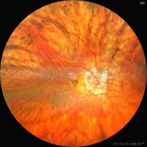 Myopic CNVM
Jul 22 2022 by T. P . VIGNESH, MBBS,MS
Fundus photograph of a 64-year-old woman with high myopia , myopic fundus and myopic CNVM.
Photographer: Bharathi Singaravel
Imaging device: Zeiss Clarus
Condition/keywords: high myopia, Myopic CNVM
-
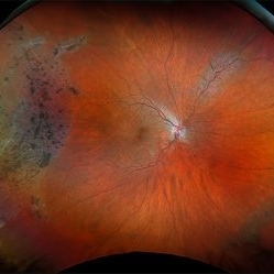
 Pigmented Paravenous Retinochoroidal Atrophy
Oct 1 2023 by Bruno DECAY, MD
Fundus photograph (autofluorescence) of a 43-year-old myopic man (left eye) Axial length: 29.32 mm
Photographer: Bruno DECAY, MD
Imaging device: iCARE EIDON confocal fundus imaging system (Ultra-Widefield Module)
Condition/keywords: Bilateral, High myopia
-
 Pigmented Paravenous Retinochoroidal Atrophy
Oct 1 2023 by Bruno DECAY, MD
Fundus photograph (autofluorescence) of a 43-year-old myopic man (right eye) Axial length: 27.67 mm
Photographer: Bruno DECAY, MD
Imaging device: iCARE EIDON confocal fundus imaging system (Ultra-Widefield Module)
Condition/keywords: Bilateral, High Myopia, Pigmented Paravenous Retinochoroidal Atrophy
-
 Pigmented Paravenous Retinochoroidal Atrophy
Oct 1 2023 by Bruno DECAY, MD
Fundus photograph of a 43-year-old myopic man (right eye) Axial length: 27.67 mm
Photographer: Bruno DECAY, MD
Imaging device: iCARE EIDON confocal fundus imaging system (Ultra-Widefield Module)
Condition/keywords: Bilateral, High myopia, Pigmented Paravenous Retinochoroidal Atrophy
-
 Retinitis Pigmentosa
Apr 9 2025 by Virginia Gebhart
35 year old female with stable sectoral RP and high myopia OU. RP has not progressed in either eye since initial visit in 2021. Will continue to observe. VA 20/20 OU
Photographer: Virginia Gebhart, Retina Consultants of Carolina
Imaging device: Optos California
Condition/keywords: high myopia, retinitis pigmentosa
-
 Submacular Hemorrhage
Mar 12 2016 by Sjakon G Tahija, MD
This a fundus photograph of a high myope who presented with a submacular hemorrhage.
Photographer: Avris Siahaan, Klinik Mata Nusantara, Jakarta, Indonesia
Condition/keywords: high myopia, spontaneous submacular hemorrhage
-
 Giant Retinal Tear
Oct 9 2012 by Audina M. Berrocal, MD FASRS
Teenager with high myopia and a GRT
Photographer: Ditte Hess CRA, BPEI
Imaging device: Fundus Camera
Condition/keywords: high myopia, retinal degeneration, retinal tear
-
 PDR; High Myopia; PRP
May 2 2019 by Carissa Hurdstrom
PDR; high myopia; PRP
Imaging device: Optos
Condition/keywords: fluorescein angiogram (FA), high myopia, pan-retinal photocoagulation (PRP), proliferative diabetic retinopathy (PDR)
-
 Inferior Rhegmatogenous Retinal Detachment with Subretinal Fibrosis
Aug 23 2012 by Gabriela Lopezcarasa Hernandez, MD
Asymptomatic 25-year-old woman with high myopia.
Photographer: Gabriela Lopezcarasa Hernandez, Hospital Angeles Lomas
Imaging device: FF4
Condition/keywords: high myopia, subretinal fibrosis
-
 Myopic CNV
Jan 11 2013 by Alex P. Hunyor, MD
Myopic macular degeneration complicated by subretinal neovascularisation, left eye.
Condition/keywords: high myopia, myopia, myopic choroidal neovascularization (CNV)
-
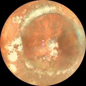
 High Myopia with Cobblestone Degeneration
Nov 5 2019 by Nichole Lewis
50-year-old female with high myopia, diffuse myopic thinning and cobblestone degeneration.
Photographer: Nichole Lewis
Imaging device: Optos
Condition/keywords: high myopia, myopia, paving stone degeneration
-
 Bilateral CNV in High Myopia
Apr 2 2019 by Gary R. Cook, MD, FACS
Right eye of a 60-year-old white female with -9D myopia, myopic maculopathy, and visible (Type 1) CNV; V.A. = 20/40.
Imaging device: Topcon VT-50
Condition/keywords: choroidal neovascular membrane (CNVM), choroidal neovascularization (CNV), high myopia, myopic degeneration, myopic fundus, pathologic myopia
-
 Bilateral CNV in High Myopia
Apr 2 2019 by Gary R. Cook, MD, FACS
Left eye of a 60-year-old white female with -9D myopia and bilateral visible (Type 1) CNV; V.A. = 20/30.
Imaging device: Topcon VT-50
Condition/keywords: choroidal neovascular membrane (CNVM), choroidal neovascularization (CNV), high myopia, myopic degeneration, myopic fundus, pathologic myopia
-
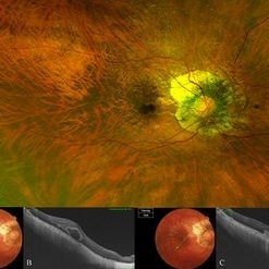 Bow-Tie Macular Hemorrhage With Cyst- Atypical Presentation of Myopic Choroidal Neovascularization
Mar 26 2021 by RUSHIK PATEL
The image of right eye of 51-year-old lady with high myopia show " Bow-Tie" macular hemorrhage (A). Optical coherence tomography (B) scan passing through hemorrhage showed intraretinal cystic lesion. During the course of intravitreal anti-VEGF injection treatment, the lesion converted into typical myopic choroidal neovascularization (C).
Photographer: Rushik Patel, Netralaya Super Speciality Eye Hospital
Condition/keywords: cyst, macular hemorrhage, myopic choroidal neovascularization (CNV)
-
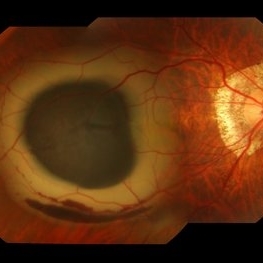
 Buckled Eye in a case of High Myopia
Jan 8 2020 by Sham Talati, DOMS
Buckled Eye in a case of high myopia.
Photographer: Dr. Sham Talati,Retina Foundation,Ahmedabad
Imaging device: Nidek Mirante
Condition/keywords: high myopia, scleral buckle
-
 Chronic Retinal Detachment in a Young Myopic Patient
Nov 6 2019 by Kamal Kishore, MD, MBBS
Chronic retinal detachment in a 27-year-old myopic female showing spontaneous reattachment in inferotemporal quadrant, and demarcation line and subretinal gliosis in superotemporal quadrant.
Photographer: Stephanie Shaver
Imaging device: Topcon 50 EX with OIS Winstation
Condition/keywords: chronic retinal detachment, high myopia
 Loading…