Search results (46 results)
-
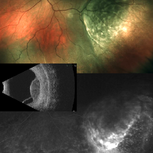
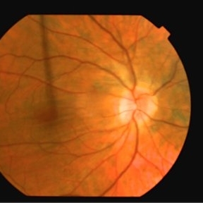
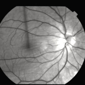
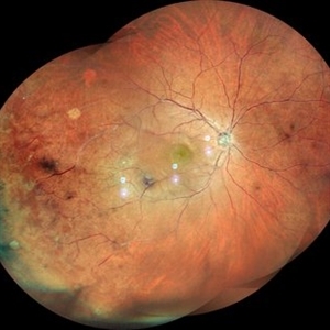
 Choroidal Melanoma with Exudative Retinal Detachment
Mar 2 2023 by Aditya S Kelkar, MS, FRCS, FASRS,FRCOphth
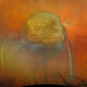
Color fundus photograph of the left eye of a 45 year old male showing choroidal melanoma with exudative retinal detachment.
Photographer: Dr. Pranali Surawase, National Institute of Ophthalmology, Pune, India.
Imaging device: Zeiss Clarus 500
Condition/keywords: choroidal mass, exudative retinal detachment, Retinal detachment
-
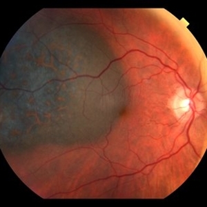
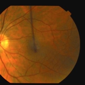
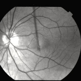
 Choroidal Melanoma with Serous Retinal Detachment
Dec 20 2024 by Daniel Davis, OCT-C
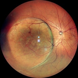
67 year old male presenting with large pigmented choroidal mass with serous retinal detachment.
Photographer: Daniel Davis, OCT-C, The Retina Institute
Imaging device: Optos California
Condition/keywords: Retina detachment
-
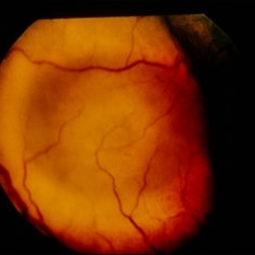
 Choroidal Mass
Sep 21 2023 by Vaidehi Sathaye
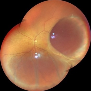
Widefield photograph of RE of a 68 year male with choroidal mass.
Photographer: Dr. Vaidehi Sathaye
Imaging device: Mirante
Condition/keywords: choroidal mass
-
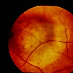
 Choroidal Melanoma
Feb 6 2025 by Virginia Gebhart
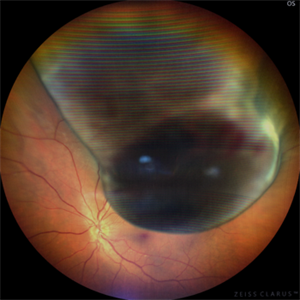
81 year old female with large pigmented collar button ciliochoroidal mass extending into the mid-vitreous cavity. Clinical exam and ultrasound finding consistent with melanoma. Due to size of tumor, pt scheduled for enucleation. CT scan of abdomen showed no evidence of metastatic disease.
Photographer: Virginia Gebhart, Retina Consultants of Carolina
Imaging device: Optos California
Condition/keywords: ciliochoroidal melanoma, collar button, melanoma
-
 Intraocular Mass With Retinal Detachment
Aug 28 2019 by Gayathri Mohan
Wide field fundus image showing an intraocular mass temporally along with a retinal detachment.
Photographer: Dr. Gayathri Mohan, Retina Foundation
Imaging device: Nidek Mirante SLO
Condition/keywords: choroidal mass
-
 Uveal Melanoma
Apr 26 2025 by Vishal Agrawal, MD, FRCS,FACS,FASRS
A 32 year-old male presented with complaints of perceiving a shadow in OS for 15-20 days. His BCVA was 20/20 OU. On Fundus examination, a large, elevated, well-defined, pigmented choroidal mass with few hemorrhages over the lesion was seen and a provisional diagnosis of uveal melanoma was made. urgent oncological consultation was recommended for further treatment.
Photographer: Dr Ayushi Gupta
Imaging device: Clarus 700
Condition/keywords: melanoma
-
 Choroidal Mass
Sep 21 2018 by Sarah Oelrich
Choroidal mass
Photographer: Sarah Oelrich CRA COT, Southeastern Retina Associates Knoxville Tn
Imaging device: OPTOS 200tx
Condition/keywords: choroidal mass
-
 Orange Pigment Overlying a Lesion Suspicious for a Choroidal Melanoma
Jan 16 2019 by John S. King, MD
76-year-old white male saw his eye doctor with a three week complaint of photopsias and a shadow in his vision. Found to have a 10.5/12.5/2.5 (medium reflectivity) pigmented, choroidal mass associated with SRF and orange pigment (hyper-autofluorescence of lipofuscin), and without drusen or halo. See photo
Photographer: Stacey Coleman
Imaging device: Topcon 50
Condition/keywords: lipofuscin, orange pigment
-
 Amelanotic Choroidal Melanoma
Jan 29 2015 by H. Michael Lambert, MD
Elevated choroidal mass.
-
 Amelanotic Choroidal Melanoma
Jan 29 2015 by H. Michael Lambert, MD
Elevated choroidal mass.
-
 Choroidal Detachment OS
Jul 5 2024 by Zach Seim
Optos Fundus Photograph of a Choroidal Detachment OS in a 75 year old male. VA at presentation was DCC HM.
Photographer: Zach Seim
Imaging device: Optos California
Condition/keywords: choroidal detachment, choroidal mass, left eye, optos, OPTOS CALIFORNIA
-
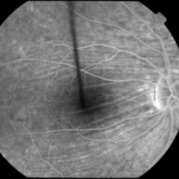
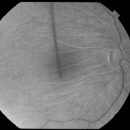
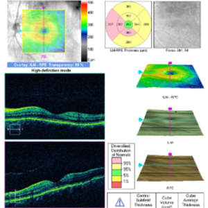
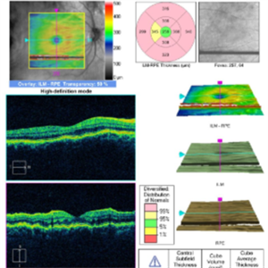
 Choroidal Folds
Nov 28 2014 by Thomas A. Ciulla, MD, MBA, FASRS
This 53-year-old man was noted to have choroidal folds right greater than left. The visual acuity was normal at 20/15. The choroidal folds are visible on OCT, especially on the vertical cuts that image across the horizontal folds. Angiography revealed staining of the folds without CNVM, choroidal mass, or optic nerve edema.
Photographer: Charlotte Harris
Condition/keywords: bilateral chorioretinal folds, choroidal folds
-
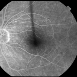
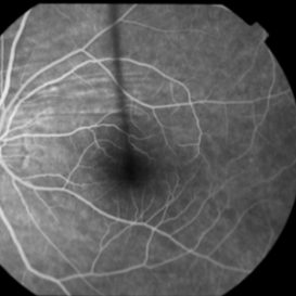
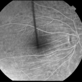
 Choroidal Folds
Nov 28 2014 by Thomas A. Ciulla, MD, MBA, FASRS
This 53-year-old man was noted to have choroidal folds right greater than left. The visual acuity was normal at 20/15. The choroidal folds are visible on OCT, especially on the vertical cuts that image across the horizontal folds. Angiography revealed staining of the folds without CNVM, choroidal mass, or optic nerve edema.
Condition/keywords: bilateral chorioretinal folds, choroidal folds
-
 Choroidal Folds
Nov 28 2014 by Thomas A. Ciulla, MD, MBA, FASRS
This 53 -year-old man was noted to have choroidal folds right greater than left. The visual acuity was normal at 20/15. The choroidal folds are visible on OCT, especially on the vertical cuts that image across the horizontal folds. Angiography revealed staining of the folds without CNVM, choroidal mass, or optic nerve edema.
Condition/keywords: bilateral chorioretinal folds, choroidal folds
-
 Choroidal Mass
Jul 14 2013 by Jason S. Calhoun
Fundus photo shows yellowish choroidal mass in the left eye.
Photographer: Jason S. Calhoun, Department of Ophthalmology, Mayo Clinic Jacksonville, Florida
Imaging device: TOPCON TRC 50-EX
Condition/keywords: choroidal nevus, indolent choroidal mass
-
 Choroidal Mass
Mar 4 2024 by ANKIT JAIN
Left eye color photo montage of 39 year old female with sub retinal mass in nasal quadrant with hemorrhages and subretinal fluid with inferior retinal detachment.
Photographer: Dr Ankit Jain
Imaging device: MIRANTE
Condition/keywords: choroidal mass
-
 Choroidal Mass
Mar 4 2024 by ANKIT JAIN
RE color photo montage of right eye of 48 year old with sub retinal hemorrhage with sub retinal fluid at level of fovea.
Photographer: Dr Ankit Jain
Imaging device: MIRANTE
Condition/keywords: macroaneurysm, retinal arterial macroaneurysm
 Loading…