Search results (113 results)
-
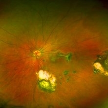
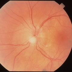
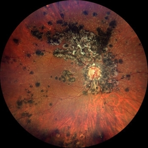 Tuberculosis-related serpiginous-like choroiditis
Nov 22 2022 by Ricardo Leitão Guerra
True color BLFI of a 60-year-old male presenting chorioretinal scars from a tuberculosis-related serpiginous-like choroiditis.
Photographer: Ricardo Leitão Guerra
Imaging device: Zeiss Clarus 700
Condition/keywords: serpiginous choroiditis, tuberculosis
-
 Chorioretinitis with Overlying Vitreous Stranding/Vitritis
Mar 23 2023 by Isaac Agranoff
Fundus photograph of a 37-year-old woman presenting with chorioretinitis with overlying vitreous stranding/vitritis that has remained unchanged for multiple years. Patient presented with irritation and blurred vision and her vision was 20/40 OD. The OCT revealed evidence of low-grade inflammation and the recommend treatment was anti-inflammatory eye drops at this time and to obtain second opinion with another physician in the office.
Photographer: Isaac Agranoff, Technician
Imaging device: Optos California
Condition/keywords: chorioretinal scar, chorioretinitis, inflammation, Optos, ultra-wide field imaging, vitritis
-
 Congenital Toxoplasmosis
Feb 2 2021 by Niloofar Piri, MD
43-year-old female with large oval chorioretinal scar in posterior pole with heavy RPE hyperplasia and history of hydrocephalus s/p VP shunt since birth. Findings are consistent with congenital toxoplasmosis.
Condition/keywords: congenital toxoplasmosis
-
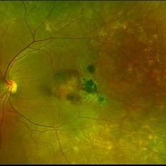
 Retinocoroiditis Inactiva Por Toxoplasmosis
Apr 28 2025 by Paulina Araujo
Fundus photography demonstrates a 2-disc-diameter chorioretinal scar in the superior temporal arcade, consistent with inactive toxoplasmic retinochoroiditis. The lesion exhibits pigmented borders and central atrophy, with adjacent splinter hemorrhages and vascular sheathing. No vitreous inflammation or active satellite lesions are present.
Photographer: Paulina D.Araujo Martínez, Asociación para Evitar la Ceguera en México I.A.P., Hospital Dr Luis Sánchez Bulnes.
Condition/keywords: toxoplasmosis chorioretinitis
-
 Choroidopathy
May 27 2020 by Jamin S. Brown, MD
Fluorescein angiography image of 28 year old female with focal chorioretinal inflammation, macular or paramacular OS. chorioretinal scar OS.
Photographer: Jeffrey Barker, Retina-Vitreous Surgeons of CNY
Condition/keywords: choroidopathy
-
 Disseminated Chorioretinitis With Unknown Etiology
Apr 5 2018 by Kim Barrett
Ultra-wide field fluorescein angiogram of a 31-year-old female with intermittent pain in her left eye. Her condition has been managed in Liberia until recently when she moved to the United States. She suffers from multiple modalities including central retinal artery occlusion, posterior synechiae of the iris, interstitial keratitis, disseminated chorioretinitis, as well as HIV. An infectious cause is high on the differential in light of her HIV status. DDx: hypertensive crisis, an embolism (? IV drug use), coagulopathy, trauma, infectious. Blood work was normal. Her current vision is 20/30 right eye and 20/400 left eye.
Photographer: Kim Barrett, COA
Imaging device: Optos
Condition/keywords: central retinal artery occlusion (CRAO), chorioretinal scar, ciliary artery sparring, disseminated chorioretinitis, HIV, left eye, optic atrophy, staining
-
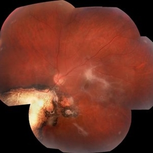
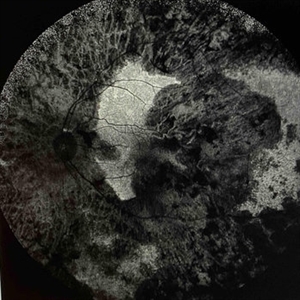 Extensive Chorioretinal Scarring With Partial Macular Sparing
Apr 22 2025 by Maxwell J Wingelaar, MD
Fundus autofluorescence of extensive chorioretinal scarring in the left eye.
Photographer: Killian Roberts
Imaging device: Heidelberg Spectralis AF
Condition/keywords: chorioretinal atrophy, chorioretinal inflammations
-
 Retinal Aneurysms
Aug 6 2025 by Korey Starkey
54 year-old patient presents with scattered peripheral aneurysms with exudates. FA was performed showing peripheral nonperfusion and aneurysms. Treated patient with PRP and focal laser to aneurysms and continued observation.
Photographer: Kore Starkey
Imaging device: Optos
Condition/keywords: aneurysm, branch retinal vein occlusion (BRVO), chorioretinal scar, circinate ring, exudates, fundus photography, lesion, Optos, retinal aneurysms
-
 Subretinal Hemorrhage with Chorioretinal Macular Scars
Sep 28 2022 by Denica Rodriguez
Ultra-widefield pseudocolor fundus photograph of a 59 year old female with Subretinal Hemorrhage with Chorioretinal Macular Scars affecting her left eye. The physician presumes the etiology is CNV from adjacent scarring/choroidal rupture. Patient has history of ocular trauma with cricket ball at age 10-12 years old. She suspects that she might have suffered choroidal rupture, which has resulted in secondary CNV and hemorrhage that we are seeing today. She recommends treatment with Eylea sample injection in a series of 4 at a 4-5 week interval. The patient's vision at the time of her appointment was Dcc20/40-2 PHNI.
Photographer: Denica Rodriguez, COA
Imaging device: Optos California
Condition/keywords: antiVEGF therapy, chorioretinal scar, choroidal neovascular membrane (CNVM), fundus photography, left eye, macular scar, Optos, peripheral drusen, pseudocolor, secondary CNV, subretinal hemorrhage, ULTRA WIDE FIELD, ultra-wide field imaging
-
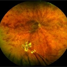
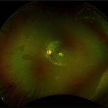
 Toxoplasma chorioretinitis 1
Jan 11 2013 by Alex P. Hunyor, MD
Toxoplasmosis 1 - chorioretinal scar from previous toxoplasma chorioretinitis. See image 2 - recurrent todo adjacent to this scar
Condition/keywords: inactive toxoplasmosis, ocular toxoplasmosis, toxoplasmosis, toxoplasmosis retinitis
-
 Toxoplasmosis
Jun 3 2017 by Gabriel Costa Andrade, PhD
Fundus photograph of an 14-year-old boy with multiple chorioretinal scars secondary to toxoplasmosis.
Photographer: Gabriel Costa de Andrade
Imaging device: Optos® California
Condition/keywords: congenital toxoplasmosis, ocular toxoplasmosis, toxoplasmosis chorioretinitis, toxoplasmosis uveitis
-
 Toxoplasmosis Associated Epiretinal Membrane
Oct 27 2016 by Gabriel Costa Andrade, PhD
Fundus photograph of a 26-year-old woman with a chorioretinal scar due to toxoplasmosis and secondary epiretinal membrane.
Photographer: Gabriel Andrade, Federal University of São Paulo, São Paulo, Brazil
Condition/keywords: epiretinal membrane (ERM), toxoplasmosis
-
 Traumatic Chorioretinal Scarring
Oct 15 2012 by Jeffrey G. Gross, MD, FASRS
Traumatic chorioretinal scarring, with less hemorrhage, 1 month later.
Condition/keywords: chorioretinal scar
-
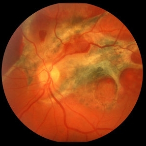
 Extensive Chorioretinal Scarring with Partial Macular Sparring
Apr 22 2025 by Maxwell J Wingelaar, MD
A multicolor photo showing chorioretinal scarring with partial macular sparing in the left eye.
Photographer: Killian Roberts
Imaging device: Heidelberg Spectralis Multicolor Photo
Condition/keywords: chorioretinal atrophy, chorioretinal inflammations
-
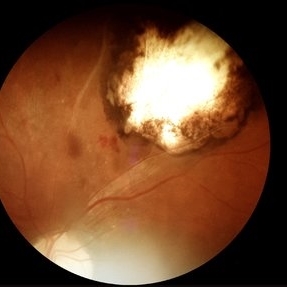
 Acute Toxoplasmosis in AIDS
Apr 8 2019 by Gary R. Cook, MD, FACS
Left eye of a white male with AIDS and an optic neuritis secondary to ocular toxoplasmosis infection. The patient had no pre-existing chorioretinal scars secondary to Toxo. An edematous optic nerve with a focus of active retinitis inferonasally, small surface hemorrhage above it, and surrounding peripapillary edema is visible.
Imaging device: Topcon VT-50
Condition/keywords: AIDS, ocular toxoplasmosis, optic neuritis, toxoplasmosis
-
---thumb.jpg/image-square;max$300,300.ImageHandler) case 2 OD
case 2 OD
Feb 14 2013 by From the Collections of Thomas M. Aaberg, MD and Thomas M. Aaberg Jr., MD
reproductions of figures 4 and 5 from the article "Ocular involvement in neonatal herpes simplex virus infection" (Hagler WS et al, Arch Opthalmol 1969;82:169-76.). Fulminating chorioretinal scarring and retinal pigmentary changes were seen in both eyes of an infant with neonatal systemic herpesvirus infection.
Condition/keywords: chorioretinal scar, neonatal herpes
-
 Central Retinal Vein Occlusion
Jul 13 2018 by Olivia Rainey
Ultra-wide field, pseudocolor montage of a patient presenting with a central retinal vein occlusion, as well as, an inferior chorioretinal scar in their right eye.
Photographer: Olivia Rainey
Imaging device: Optos
Condition/keywords: central retinal vein occlusion (CRVO), chorioretinal scar, montage, Optos, pseudocolor, ultra-wide field imaging
-
 Chorio Retinal Scar
Feb 19 2024 by Sanauddin Samejo , Diploma (Ophthalmic Technician Training Course)
A patient came in to Clinic of Dr Madhav Rao (VR Surgeon)
Photographer: Sanauddin Samejo, Burjeel Hospital, Abu Dhabi, UAE.
Imaging device: Optos Silverstone
Condition/keywords: chorioretinal scar
-
 Chorioretinal Macula Scar (Macula View)
May 12 2025 by Briana Hernandez
Zoomed in Macular View of Chorioretinal Macular Scar in 9-year-old female patient.
Photographer: Briana Hernandez, Hilton Head Retina Insitute
Imaging device: Optos
Condition/keywords: chorioretinal scar
-
 Chorioretinal Macula Scar (Ultrawide View)
May 12 2025 by Briana Hernandez
Ultra wide Optos image of Chorioretinal Macular Scar in 9-year-old female patient.
Photographer: Briana Hernandez, Hilton Head Retina Institute
Imaging device: Optos
Condition/keywords: chorioretinal scar, macular scar, ultra-wide field imaging
-
 Chorioretinal Scar
Feb 19 2024 by Sanauddin Samejo , Diploma (Ophthalmic Technician Training Course)
A patient came in to the clinic of Dr Madhav Rao (VR Surgeon).
Photographer: Sanauddin Samejo, Burjeel Hospital, Abu Dhabi, UAE.
Imaging device: Optos Silverstone
Condition/keywords: retinal scar
-
 Chorioretinal Scar
May 16 2017 by Olivia Rainey
Fundus photograph of an 17-year-old male with a macular scar affecting his right eye secondary to exudation from Coats disease.
Photographer: Olivia Rainey
Imaging device: Topcon 50dx
Condition/keywords: 20 degrees, chorioretinal scar, Coats' disease, color fundus photograph, color photo, fundus photograph
-
 Chorioretinal Scar
Apr 1 2016 by Nichole Lewis
Chorioretinal scar.
Photographer: Nichole Lewis - Pennsylvania Retina Specialists, Camp Hill, PA
Condition/keywords: chorioretinal scar
-
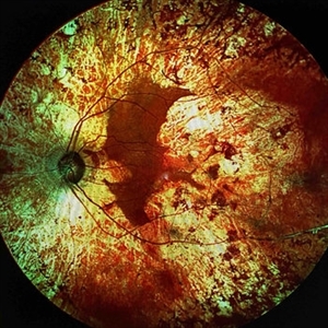
---thumb.jpg/image-square;max$300,300.ImageHandler) Chorioretinal Scarring
Chorioretinal Scarring
Feb 15 2013 by From the Collections of Thomas M. Aaberg, MD and Thomas M. Aaberg Jr., MD
Color fundus photograph showing chorioretinal scarring consistent with prior retinal laser photocoagulation to areas of peripheral retinal nonperfusion.
Condition/keywords: laser scarring, peripheral retinal nonperfusion
-
 Chorioretinal Scars with Subretinal Fibrosis and an old Retinal Detachment
May 3 2018 by Nichole Lewis
Chorioretinal scars with subretinal fibrosis and an old retinal detachment.
Photographer: Nichole Lewis
Condition/keywords: chorioretinal scar, chronic retinal detachment, subretinal fibrosis
 Loading…