Search results (5 results)
-
 Slide 1-20
Feb 19 2019 by Lancaster Course in Ophthalmology
Langhans giant cell with peripheral nuclei in the reaction around a pool of fat, from a chalazion of the lid. (H&E stain)
Condition/keywords: chalazion, Langhans giant cell, nuclei
-
 Slide 3-27
Feb 20 2019 by Lancaster Course in Ophthalmology
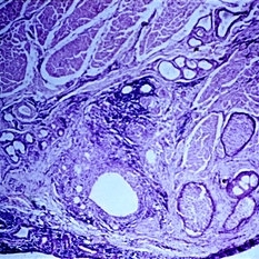
Low-power view of chalazion. Note the proximity of the inflammation to the meibomian glands.
Condition/keywords: chalazion, inflammation, meibomian glands
-
 Slide 3-28
Feb 20 2019 by Lancaster Course in Ophthalmology
Area of chalazion showing lipid droplets represented by empty spaces and predominantly chronic inflammation.
Condition/keywords: chalazion, chronic inflammation, lipid
-
 Slide 3-29
Feb 20 2019 by Lancaster Course in Ophthalmology
Pyogenic granuloma which frequently follows chalazion ( x65). In spite of its name it is neither pyogenic nor a granuloma. Note the profusion of newly formed blood vessels, inflammatory cells, and connective tissue.
Condition/keywords: chalazion, granuloma
-
 Slide 5-8
Feb 20 2019 by Lancaster Course in Ophthalmology
Small chalazion in situ in the lid. Lipogranulomatous reaction around large and small clear spaces (pools of dissolved fat) from a single obstructed meibomian lobule.
Condition/keywords: chalazion, lipogranulomatous reaction, meibomian lobule, situ
 Loading…