Search results (102 results)
-
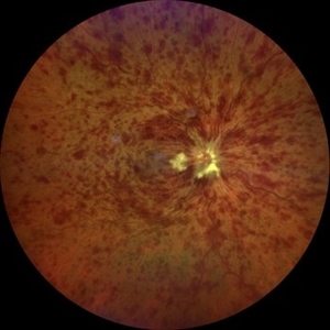 Central Retinal Vein Occlusion associated with disc edema
Oct 19 2023 by Gabriel Costa Andrade, PhD
53-year-old woman with an acute CRVO. The patient has a history of breast cancer undergoing treatment with systemic chemotherapy. Notice the peripapillary cotton wool spots, superficial flame shaped hemorrhages and deeper dot and blot hemorrhages in all 4 quadrants.
Photographer: Gabriel Andrade
Condition/keywords: central retinal vein occlusion (CRVO), macular edema, Retina
-
 Breast Cancer Metastatic Lesion to Choroid
Oct 16 2012 by Jeffrey G. Gross, MD, FASRS
Breast cancer, metastatic lesion to choroid.
Condition/keywords: choroid, metastatic lesion
-
 Metastatic Breast Cancer / Iris & Choroid
Mar 25 2014 by David Callanan, MD
45-year-old female, metastatic breast CA / iris & choroid.
Condition/keywords: choroid, iris
-
 Metastatic Breast Carcinoma
Jan 21 2021 by Jamin S. Brown, MD
This anterior segment photograph was taken with a smartphone camera attached to a regular Haag Streit slit lamp ocular demonstrates unusual clustering of white cells on the posterior surface of the intraocular lens. The clinical diagnosis is metastatic breast carcinoma to the vitreous, which is very rare.
Photographer: Stefanie Palmer CRA, Retina Vitreous Surgeons of CNY
Imaging device: Cell phone camera
Condition/keywords: anterior segment, breast cancer, cell phone camera, slit lamp photo
-
 Tamoxifen Retinopathy- FAF
Aug 30 2012 by Young Hee Yoon, MD, PhD
Fundus autofluorescence (FAF) of an 58-year-old woman with a bilateral tamoxifen maculopathy. She had taken tamoxifen for 24 months due to breast cancer. In spite of discontinuation 2 years ago, her macula remained unchanged. Her best-corrected visual acuity was 20/50 in the right and 20/100 in the left.
Photographer: Soo Hyun Cho, Asan Medical Center
Imaging device: Heidelberg HRA II
Condition/keywords: drug toxicity, toxic maculopathy
-
 Tamoxifen Retinopathy- OCT
Aug 30 2012 by Young Hee Yoon, MD, PhD
OCT image of an 58-year-old woman with a bilateral tamoxifen maculopathy. She had taken tamoxifen for 24 months due to breast cancer. In spite of discontinuation 2 years ago, her macula remained unchanged. Her best-corrected visual acuity was 20/50 in the right and 20/100 in the left.
Photographer: Soon Tae Kim, Asan Medical Center
Imaging device: Zeiss cirrus HD-OCT 4000
Condition/keywords: drug toxicity, toxic maculopathy
-
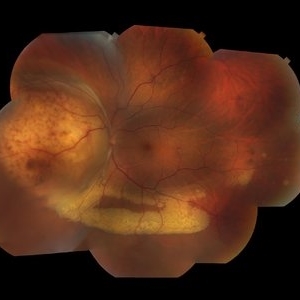 Ocular Metastasis of Breast Cancer
Mar 13 2018 by Olivia Rainey
Color fundus montage of a 45-year-old female presenting with ocular metastasis affecting her left eye. She had been treated for pneumonia, had progressive lumbar back pain, and a 29 pound weight loss recently. She reported that she had a breast lump and a mammogram, but had not been provided the results. After she was sent to oncology, it was confirmed that she has widely metastatic breast cancer and tumors throughout the brain. The oncology team felt she could not wait weeks for her chemotherapy to start and consequently decided to do whole brain radiation and treat the affected eye just posterior to the lens.
Photographer: Olivia Rainey
Imaging device: Topcon DX50
Condition/keywords: choroidal metastasis, color fundus photograph, exudative detachment, left eye, lipid exudation, montage
-
 Tamoxifen Retinopathy- Fundus photo
Aug 30 2012 by Young Hee Yoon, MD, PhD
Fundus photograph of an 58-year-old woman with a bilateral tamoxifen maculopathy. She had taken tamoxifen for 24 months due to breast cancer. In spite of discontinuation 2 years ago, her macula remained unchanged. Her best-corrected visual acuity was 20/50 in the right and 20/100 in the left.
Photographer: Ji Hee Kim, Asan Medical Center
Imaging device: Canon CR-DGI
Condition/keywords: drug toxicity, toxic maculopathy
-
 Breast cancer metastatic to choroid
Jul 13 2021 by Odette M. Houghton, MD
Montage photograph of a 59-year-old female with a choroidal tumor secondary to metastatic breast cancer.
Photographer: David Saiz COT, Mayo Clinic Arizona
Imaging device: Topcon
Condition/keywords: breast cancer, metastatic cancer, metastatic lesion
-
---thumb.jpg/image-square;max$300,300.ImageHandler) Tamoxifen Retinopathy- OCT
Tamoxifen Retinopathy- OCT
Aug 30 2012 by Young Hee Yoon, MD, PhD
OCT image of an 58-year-old woman with a bilateral tamoxifen maculopathy. She had taken tamoxifen for 24 months due to breast cancer. In spite of discontinuation 2 years ago, her macula remained unchanged. Her best-corrected visual acuity was 20/50 in the right and 20/100 in the left.
Photographer: Soon Tae Kim, Asan Medical Center
Imaging device: Heidelberg Spectralis
Condition/keywords: drug toxicity
-
 Bilateral Metastatic Lesions Secondary to Breast Cancer
Feb 18 2014 by Gabriela Lopezcarasa Hernandez, MD
Asymptomatic 44-year-old woman who went to a general exam to the ophthalmologist.
Photographer: Araceli Rojas Arriaga, Hospital Angeles Lomas, Mexico
Imaging device: ZEISS FF4
Condition/keywords: metastatic lesion
-
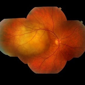
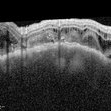 Breast cancer metastatic to choroid
Jul 13 2021 by Odette M. Houghton, MD
EDI-OCT image of a 59-year-old female with a choroidal tumor secondary to metastatic breast cancer.
Photographer: David Saiz COT, Mayo Clinic Arizona
Imaging device: Heidelberg Spectralis
Condition/keywords: breast cancer, choroidal metastasis, metastatic lesion
-
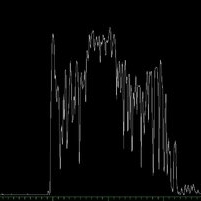 Breast cancer metastatic to choroid
Jul 13 2021 by Odette M. Houghton, MD
A-scan image of a 59-year-old female with a choroidal tumor secondary to metastatic breast cancer.
Photographer: Christina Carpenter COA, ROUB, OSC, Mayo Clinic Arizona
Imaging device: Ellex
Condition/keywords: a-scan image, breast cancer, metastatic cancer
-
 Breast cancer metastatic to choroid
Jul 13 2021 by Odette M. Houghton, MD
B-scan image of a 59-year-old female with a choroidal tumor secondary to metastatic breast cancer.
Photographer: Christina Carpenter COA, ROUB, OSC, Mayo Clinic Arizona
Imaging device: Ellex
Condition/keywords: B scan ultrasound, breast cancer, metastatic cancer
-
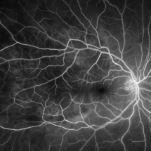 Breast cancer metastatic to choroid
Jul 13 2021 by Odette M. Houghton, MD
Arteriovenous phase fluorescein angiogram of a 59-year-old female with a choroidal tumor secondary to metastatic breast cancer.
Photographer: David Saiz COT, Mayo Clinic Arizona
Imaging device: Optos California
Condition/keywords: breast cancer, choroidal metastasis, metastatic lesion
-
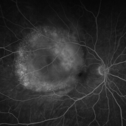 Breast cancer metastatic to choroid
Jul 13 2021 by Odette M. Houghton, MD
Late phase fluorescein angiogram of a 59-year-old female with a choroidal tumor secondary to metastatic breast cancer.
Photographer: David Saiz COT, Mayo Clinic Arizona
Imaging device: Optos California
Condition/keywords: breast cancer, FA late phase, metastatic cancer
-
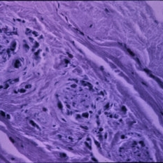 Breast Cancer Pathology Slide
-
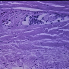 Breast Cancer Pathology Slide
-
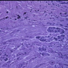 Breast Cancer Pathology Slide
-
 Breast Cancer Pathology Slide
 Loading…