Search results (222 results)
-
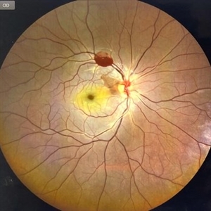
 Post Traumatic Optic Nerve Head Avulsion
Nov 18 2017 by Vishal Agrawal, MD, FRCS,FACS,FASRS
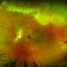
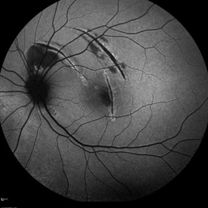
Right eye fundus picture of a 24-year-old male patient who suffered blunt trauma 7 days back with a wooden stick . He presented with NLP vision with a non reacting dilated pupil. Fundus montage picture shows ONH avulsion,CRAO,peripapillary resolving hemorrhages and cicatricial tissue at the edge.
Photographer: Vishal Agrawal, MD, SMS Medical College, Jaipur, India
Imaging device: Zeiss 524
Condition/keywords: avulsion, central retinal artery occlusion (CRAO)
-
 Commotio Post-Blunt Trauma
Apr 3 2018 by Paulo Bueno
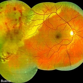
Fundus photograph of an 35-year-old man with commotio retinae after indoor soccer ball blunt trauma.
Photographer: Paulo Bueno, Taubaté, Brazil.
Imaging device: Zeiss Visucam
Condition/keywords: blunt trauma, commotio retinae
-
 Hourglass in an Eye
Apr 22 2025 by KRISHNENDU NANDI, MS
A twenty-five-year-young male presented with a decrease in vision in the right eye following a blunt trauma with a football. On examination the BCVA in the right eye was CFCF and the left eye was 6/6, N6. The anterior segment was within normal limits. AT was 12 and 10 mm of Hg in the right and left eyes, respectively. Fundus examination reveals subhyaloid haemorrhage in the right eye with an attached retina. The fundus of the left eye was within normal limits. YAG laser hyaloidotomy was done with an energy of 2 mJ in the right eye. After 3 weeks the BCVA in the right eye improved to 6/9, N6.
Photographer: Dr. Krishnendu Nandi
Imaging device: Topcon
Condition/keywords: Trauma, YAG HYALOIDOTOMY, Young Male
-
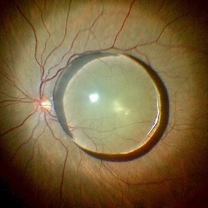
 Intraocular lens luxated to the vitreous cavity
Jun 24 2023 by Mariam Cernichiaro-Espinosa, MD
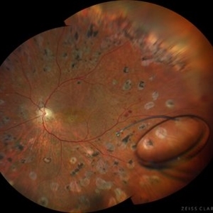
Three-piece intraocular lens luxated to the vitreous cavity in a patient with photocoagulated diabetic retinopathy after blunt trauma.
Photographer: Mariam Cernichiaro-Espinosa, Asociación para Evitar la Ceguera en México, I.A.P. Mexico City, Mexico.
Imaging device: Zeiss Clarus
Condition/keywords: diabetic retinopathy, intraocular lense in vitreous, lens luxation
-
 A Large Break at the Posterior Pole With RD With PVR (S/p Old Blunt Trauma)
Jan 16 2025 by Anand Temkar
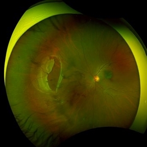
Right eye widefield fundus color photo of a 10 year old kid who noticed diminution of vision in right eye since a month. We can see the large break at the posterior pole with rolled up margins associated with retinal detachment and PVR changes.
Photographer: Dr.Anand Temkar- Retina Foundation, Ahmedabad
Imaging device: Mirante
Condition/keywords: posterior pole break, proliferative vitreoretinopathy (PVR), Retinal Detachment
-
 Dropped Crystalline Lens
Mar 8 2019 by Abdulaziz A. Alshamrani, MD
A 15-year-old female with congenital glaucoma complaining of acute diminution of vision after a blunt trauma.
Condition/keywords: crystalline lens, dropped nucleus, ora serrata
-
 Large Retinal Tear from a Shuttlecock Injury
Oct 11 2024 by Ahmad B. Tarabishy, MD
27 year old woman presenting with floaters and a shadow in her temporal visual field OS. Approximately one week earlier, she was injured in her left eye by a shuttlecock while playing badminton. Fundus exam reveals mild vitreous hemorrhage and a large retinal tear with a small cuff of surrounding SRF.
Photographer: Angela Rico, M.D.
Imaging device: Optos
Condition/keywords: blunt trauma, ocular trauma, retinal tear
-
 Massive Commotio Retinae
Oct 20 2020 by Veronika Yehezkeli
Fundus photograph of a 24-year-old male, made after blunt trauma with a plastic bottle. Note massive commotio retinae and preretinal hemorrhages in the contralateral to trauma area.
Photographer: Veronika Yehezkeli, Meir medical center, Israel
Condition/keywords: blunt trauma, commotio retinae, preretinal hemorrhage, trauma
-
 A Large Break at the Posterior Pole With RD With PVR (S/p Old Blunt Trauma)
Jan 16 2025 by Anand Temkar
Right eye central fundus color photo of a 10 year old kid who noticed diminution of vision in right eye since a month. We can see the large break at the posterior pole with rolled up margins associated with retinal detachment and PVR changes.
Photographer: Dr.Anand Temkar- Retina Foundation, Ahmedabad
Imaging device: Mirante
Condition/keywords: Posterior pole break, proliferative vitreoretinopathy (PVR), Retinal Detachment
-
 Choroidal Rupture
Apr 7 2025 by Ramses Rosales-Diaz
Autofluorescence image of a 39-year-old female patient who sustained blunt ocular trauma resulting in three choroidal ruptures.
Photographer: Ramses Rosales-Diaz, Asociación Para Evitar la Ceguera en México I.A.P., Mexico City
Imaging device: Heidelberg Spectralis
Condition/keywords: blunt trauma, Choroidal Rupture
-
 Choroidal rupture and peripapillary hemorrhage - FA
Jan 26 2013 by Roy Schwartz, MD
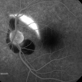
A 36-year-old male presented to the ER after blunt trauma to his left eye. On FA a chroidal rupture (hyperfluorescent area) was seen as well as peripapillary hemorrhage (hypofluorescent).
Photographer: Galit Yair-Pur
Condition/keywords: choroidal rupture, peripapillary hemorrhage
-
 Dislocated Intraocular Lens (IOL)
Aug 2 2019 by JEFFERSON R SOUSA, Tecg.º (Biomedical Systems Technology)
A 53-year-old male patient suffered blunt trauma 15 days after cataract surgery. Note total dislocation of the intraocular lens. No glass reaction.
Photographer: JEFFERSON R SOUSA - Study Center and Ophthalmological Research Dr. Andre M V Gomes, Institute Dr. Suel Abujamra São Paulo-Brazil
Imaging device: Topcon TRC-50 DX, Imaginet 4.0, angle de 50 graus. Flash 18w-s
Condition/keywords: dislocated intraocular lens (IOL)
-
 Hypotonous Maculopathy
Jul 12 2022 by Akansha Sharma
FUNDUS PHOTOGRAPH OF A 37 YEAR OLD MALE WITH HISTORY OF BLUNT TRAUMA WITH TENNIS BALL PRESENTING WITH HYPOTONOUS MACULOPATHY
Photographer: Dr. Akansha Sharma-Retina Foundation, Ahmedabad
Condition/keywords: hypotonous retinopathy
-
 Iridodialysis and Cyclodialysis, UBM
Jun 29 2018 by Gareth Lema, MD, PhD
Traumatic cataract and iridodialysis after blunt trauma. The UBM image also shows a cyclodialysis.
Photographer: Peter Buch, Ross Eye Institute, University at Buffalo Jacobs School of Medicine, Buffalo, NY
Condition/keywords: cyclodialysis, iridodialysis, traumatic cataract, ultrasound
-
 Juxtafoveal Choroidal Neovascularization Secondary to Choroidal Rupture
Aug 30 2012 by Young Hee Yoon, MD, PhD
Fluorescence Angiography (FA) image of a 14-year-old boy with a history of blunt trauma to his left eye 9 months ago. Best-corrected visual acuity remained at 20/30.
Photographer: Heon Eui Hong, Asan Medical Center
Imaging device: HHeidelberg HRA II/ version 1.7.0.0
Condition/keywords: choroidal rupture, juxtafoveal choroidal neovascularization (CNV)
-
 Luxated lens to anterior segment
Sep 7 2022 by JEFFERSON R SOUSA, Tecg.º (Biomedical Systems Technology)
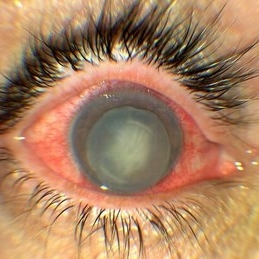
Patient 61 years old, Female, subta low vision after blunt trauma. In the anterior segment photograph, the presence of a lens in the anterior chamber is observed. In the previous follow-up OCT, the disorganization of this follow-up is clear. Above all, the documentation of these cases is essential for future decisions. This patient was urgently referred for a surgical procedure, mainly to control the intraocular pressure, which was at 60 IOP.
Photographer: JEFFERSON ROCHA DE SOUSA - Retinal Department at Instituto Dr. Suel Abujamra Sao Paulo-Brazil.
Imaging device: Clarus 700 - Zeiss,
Condition/keywords: lens luxation, Luxated lens to anterior segment, subluxation of lens
-
 OCT - Traumatic Full Thickness Macular Hole
Feb 6 2019 by awaneesh m upadhyay, MBBS, DNB
Right eye OCT image of an 8-year-old boy shows full thickness macular hole following blunt trauma of 1 week duration.
Photographer: Awaneesh Upadhyay
Imaging device: HEILDERBERG - HRA
Condition/keywords: traumatic macular hole
-
 Post traumatic dislocated crystalline lens
Jul 10 2022 by Vishal Agrawal, MD, FRCS,FACS,FASRS
Intra operative picture of a 70 year old female who presented with a history of blunt trauma and vision loss .
Photographer: Vishal Agrawal MD
Imaging device: SONY PMW 10 MD
Condition/keywords: dislocated crystalline lens, retina, trauma
-
 Traumatic Cataract and Iridodialysis, Retroillumination
Jun 29 2018 by Gareth Lema, MD, PhD
Traumatic cataract and iridodialysis after blunt trauma. Retroillumination better captured the findings that straight fundus photography.
Photographer: Peter Buch, Ross Eye Institute, University at Buffalo Jacobs School of Medicine, Buffalo, NY
Condition/keywords: iridodialysis, traumatic cataract
-
 Traumatic CRAO with Cilioretinal Artery Sparing
Sep 10 2024 by KRISHNENDU NANDI, MS
A 25 year-old male presented with dimness of vision in the right eye for the last 3 days following blunt trauma. The BCVA of the right eye was CF close to the face and left eye was 6/6, N6 in Snellen’s chart. On examination the retina showed CRAO with cherry red spot and sparing of cilioretinal artery circulation. Traumatic subhyaloid haemorrhage also noted at supero-temporal arcade.
Photographer: Dr Krishnendu Nandi
Condition/keywords: CRAO, subhyaloid hemorrhage, Trauma
-
 Traumatic macular hole
Dec 19 2012 by Eric A. Postel, MD
Color fundus photograph of a young male with a traumatic macular hole
Condition/keywords: blunt trauma, macular hole
-
 Traumatic Macular Hole
Aug 23 2012 by Gerardo Garcia-Aguirre, MD
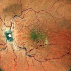
Fundus photograph of a large macular hole with an area of pigment migration secondary to blunt trauma.
Photographer: Noemí Hernández, Asociación para Evitar la Ceguera en México
Imaging device: Zeiss FF4
Condition/keywords: deformity, macular hole
-
 Subconjuntival IOL After Blunt Trauma
Jun 27 2018 by Gabriel Costa Andrade, PhD
A 73-year-old male patient was referred to our ophthalmic emergency department with complaints of redness, pain, and diminution of vision in his left eye, after fall from height. The patient underwent small incision cataract surgery with polymethylmethacrylate (PMMA) IOL implantation in both the eyes 15 years back through superior sclerocorneal incision under local anesthesia. His best-corrected visual acuity was perception of light in the left eye. Ophthalmic examination using slit lamp biomicroscopy of the left eye revealed diffuse subconjunctival hemorrhage with no conjunctival laceration and inferior bulbar conjunctiva showed traumatic pseudophacocele with a sign “golden half ring,” suggesting the presence of PCIOL in subconjunctival space.There was total hyphema obscuring the view of rest of the ocular structures in his left eye.
Photographer: Gabriel Andrade, RETINA CLINIC, São Paulo, BRAZIL
Condition/keywords: dislocated intraocular lens (IOL), trauma
-
 Traumatic Lens Drop in Vitreous
Dec 15 2020 by Manish Nagpal, MD, FRCS (UK), FASRS
Patient had come to us status post blunt trauma with the lens dislocated in inferior vitreous.
Photographer: Gayathri Mohan, Retina Fellow, Retina Foundation, Ahmedabad, India
Imaging device: Mirante CSLO
Condition/keywords: dropped nucleus, lens dislocation, traumatic cataract
-
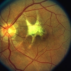 Traumatic Mac Scar
May 2 2013 by Henry J. Kaplan, MD
Traumatic stellate shaped subretinal macular scar.
Condition/keywords: blunt trauma, macular scar
 Loading…