Search results (49 results)
-
---thumb.jpg/image-square;max$300,300.ImageHandler) Birdshot Case #1 OD IVFA
Birdshot Case #1 OD IVFA
May 1 2013 by Armando L. Oliver, MD
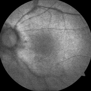
64-year-old Puerto Rican woman consulted due to the presence of 1+ vitreous cells. The fundus examination revealed orange to yellow lesions dispersing from the disk. Work-up revealed she was HLA-A29 positive and the suspected diagnosis of Birdshot Chorioretinopathy was made. Chest X-Ray, FTA-Abs and RPR were negative.
Photographer: Moises Castro, Instituto de Ojos y Piel, Carolina, PR
Imaging device: Zeiss, Visucam NM/FA
Condition/keywords: birdshot, birdshot chorioretinopathy, birdshot retinochoroidopathy
-
 Birdshot: a View From the Outside
Nov 3 2019 by Julia Farah, MD
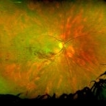
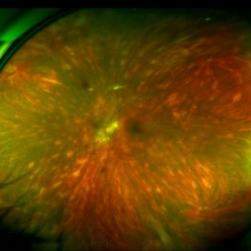
61-year-old female presented with classic birdshot chorioretinopathy.
Photographer: Peter Guingab
Imaging device: Optos California
Condition/keywords: birdshot choroidopathy, uveitis, white dot syndrome
-
 Fluocinolone Implant
Sep 12 2012 by Pauline T Merrill, MD, FASRS
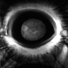
Slit lamp photograph of a Retisert fluocinolone implant in a 52-year-old male with birdshot chorioretinopathy.
Photographer: Pauline Merrill, MD
Imaging device: iPhone photo through slit lamp
Condition/keywords: birdshot, chronic uveitis, fluocinolone implant
-
 Birdshot Chorioretinopathy FA with Patchy Choroidal Filling Left Eye
Oct 9 2012 by Jeffrey G. Gross, MD, FASRS
Birdshot chorioretinopathy, FA, with patchy choroidal filling, left eye.
Condition/keywords: birdshot, chorioretinopathy, left eye, patchy choroidal filling
-
 CRVO on Birdshot
Aug 14 2017 by Mark E Kleinman, MD
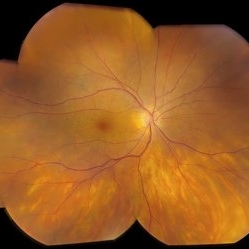
Ultra-widefield color fundus image at presentation of a 45-year-old woman with CRVO and Birdshot chorioretinopathy which was confirmed A29 with HLA typing.
Photographer: Deborah McDonald, University of Kentucky, Advanced Eye Care
Imaging device: Optos California
Condition/keywords: birdshot chorioretinopathy, central retinal vein occlusion (CRVO)
-
 Birdshot
Jul 14 2013 by Jason S. Calhoun
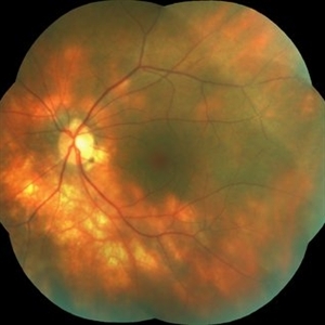
Follow up on patient with birdshot chorioretinopathy in both eyes. Posterior uveitis in both eyes no changes in inflammation.
Photographer: Jason S. Calhoun, Department of Ophthalmology, Mayo Clinic Jacksonville, Florida
Imaging device: TOPCON TRC 50-EX
Condition/keywords: birdshot chorioretinopathy
-
---thumb.jpg/image-square;max$300,300.ImageHandler) Birdshot Case #1 OD Color
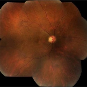
Birdshot Case #1 OD Color
May 1 2013 by Armando L. Oliver, MD
64-year-old Puerto Rican woman consulted due to the presence of 1+ vitreous cells. The fundus examination revealed orange to yellow lesions dispersing from the disk. Work-up revealed she was HLA-A29 positive and the suspected diagnosis of Birdshot Chorioretinopathy was made. Chest X-Ray, FTA-Abs and RPR were negative.
Photographer: Moises Castro, Instituto de Ojos y Piel, Carolina, PR
Imaging device: Zeiss, Visucam NM/FA
Condition/keywords: birdshot, birdshot chorioretinopathy, birdshot retinochoroidopathy
-
---thumb.jpg/image-square;max$300,300.ImageHandler) Birdshot Case #1 OD FAF
Birdshot Case #1 OD FAF
May 1 2013 by Armando L. Oliver, MD
64-year-old Puerto Rican woman consulted due to the presence of 1+ vitreous cells. The fundus examination revealed orange to yellow lesions dispersing from the disk. Work-up revealed she was HLA-A29 positive and the suspected diagnosis of Birdshot Chorioretinopathy was made. Chest X-Ray, FTA-Abs and RPR were negative.
Photographer: Moises Castro, Instituto de Ojos y Piel, Carolina, PR
Imaging device: Zeiss, Visucam NM/FA
Condition/keywords: birdshot, birdshot chorioretinopathy, birdshot retinochoroidopathy
-
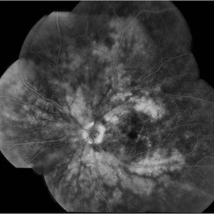
---thumb.jpg/image-square;max$300,300.ImageHandler) Birdshot Case #1 OD IVFA
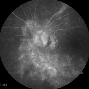
Birdshot Case #1 OD IVFA
May 1 2013 by Armando L. Oliver, MD
64-year-old Puerto Rican woman consulted due to the presence of 1+ vitreous cells. The fundus examination revealed orange to yellow lesions dispersing from the disk. Work-up revealed she was HLA-A29 positive and the suspected diagnosis of Birdshot Chorioretinopathy was made. Chest X-Ray, FTA-Abs and RPR were negative.
Photographer: Moises Castro, Instituto de Ojos y Piel, Carolina, PR
Imaging device: Zeiss, Visucam NM/FA
Condition/keywords: birdshot, birdshot chorioretinopathy, birdshot retinochoroidopathy
-
 Birdshot Case #1 OS Color
May 1 2013 by Armando L. Oliver, MD
64-year-old Puerto Rican woman consulted due to the presence of 1+ vitreous cells. The fundus examination revealed orange to yellow lesions dispersing from the disk. Work-up revealed she was HLA-A29 positive and the suspected diagnosis of Birdshot Chorioretinopathy was made. Chest X-Ray, FTA-Abs and RPR were negative.
Photographer: Moises Castro, Instituto de Ojos y Piel, Carolina, PR
Imaging device: Zeiss, Visucam NM/FA
Condition/keywords: birdshot, birdshot chorioretinopathy, birdshot retinochoroidopathy
-
---thumb.jpg/image-square;max$300,300.ImageHandler) Birdshot Case #1 OS Color
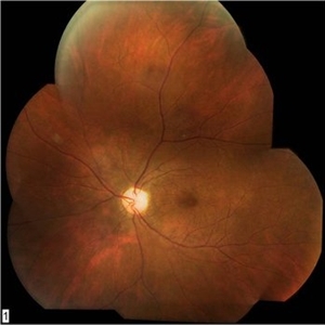
Birdshot Case #1 OS Color
May 1 2013 by Armando L. Oliver, MD
64-year-old Puerto Rican woman consulted due to the presence of 1+ vitreous cells. The fundus examination revealed orange to yellow lesions dispersing from the disk. Work-up revealed she was HLA-A29 positive and the suspected diagnosis of Birdshot Chorioretinopathy was made. Chest X-Ray, FTA-Abs and RPR were negative.
Photographer: Moises Castro, Instituto de Ojos y Piel, Carolina, PR
Imaging device: Zeiss, Visucam NM/FA
Condition/keywords: birdshot, birdshot chorioretinopathy, birdshot retinochoroidopathy
-
---thumb.jpg/image-square;max$300,300.ImageHandler) Birdshot Case #1 OS FAF
Birdshot Case #1 OS FAF
May 1 2013 by Armando L. Oliver, MD
64-year-old Puerto Rican woman consulted due to the presence of 1+ vitreous cells. The fundus examination revealed orange to yellow lesions dispersing from the disk. Work-up revealed she was HLA-A29 positive and the suspected diagnosis of Birdshot Chorioretinopathy was made. Chest X-Ray, FTA-Abs and RPR were negative.
Photographer: Moises Castro, Instituto de Ojos y Piel, Carolina, PR
Imaging device: Zeiss Visucam NM/FA
Condition/keywords: birdshot, birdshot chorioretinopathy, birdshot retinochoroidopathy
-
---thumb.jpg/image-square;max$300,300.ImageHandler) Birdshot Case #1 OS IVFA
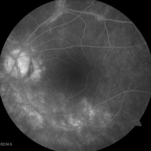
Birdshot Case #1 OS IVFA
May 1 2013 by Armando L. Oliver, MD
64-year-old Puerto Rican woman consulted due to the presence of 1+ vitreous cells. The fundus examination revealed orange to yellow lesions dispersing from the disk. Work-up revealed she was HLA-A29 positive and the suspected diagnosis of Birdshot Chorioretinopathy was made. Chest X-Ray, FTA-Abs and RPR were negative.
Photographer: Moises Castro, Instituto de Ojos y Piel, Carolina, PR
Imaging device: Zeiss, Visucam NM/FA
Condition/keywords: birdshot, birdshot retinochoroidopathy
-
 Birdshot Case #1 OS IVFA
May 1 2013 by Armando L. Oliver, MD
64-year-old Puerto Rican woman consulted due to the presence of 1+ vitreous cells. The fundus examination revealed orange to yellow lesions dispersing from the disk. Work-up revealed she was HLA-A29 positive and the suspected diagnosis of Birdshot Chorioretinopathy was made. Chest X-Ray, FTA-Abs and RPR were negative.
Photographer: Moises Castro, Instituto de Ojos y Piel, Carolina, PR
Imaging device: Zeiss, Visucam NM/FA
Condition/keywords: birdshot, birdshot chorioretinopathy, birdshot retinochoroidopathy
-
 Birdshot Case #1 OS IVFA
May 1 2013 by Armando L. Oliver, MD
64-year-old Puerto Rican woman consulted due to the presence of 1+ vitreous cells. The fundus examination revealed orange to yellow lessions dispersing from the disk. Work-up revealed she was HLA-A29 positive and the suspected diagnosis of Birdshot Chorioretinopathy was made. Chest X-Ray, FTA-Abs and RPR were negative.
Photographer: Moises Castro, Instituto de Ojos y Piel, Carolina, PR
Imaging device: Zeiss, Visucam NM/FA
Condition/keywords: birdshot, birdshot chorioretinopathy, birdshot retinochoroidopathy
-
 Birdshot Case # 2, Color OD
May 1 2013 by Armando L. Oliver, MD
57-year-old Puerto Rican woman presented for evaluation given history of chronic macular edema. Fundus examination revealed oval orange lesions dispersing from the disk. The patient has history of Type 2 DM. Work-up revealed that she was HLA-A29 positive. The patient had a negative CXR, FTA-Abs and RPR.
Photographer: Hector Mendez Caratini, Hyde Park Ophthalmology Group, San Juan, PR
Imaging device: Topcon TRC 50DX
Condition/keywords: birdshot, birdshot chorioretinopathy, birdshot retinochoroidopathy
-
 Birdshot Case # 2, Color OS
May 1 2013 by Armando L. Oliver, MD
57-year-old Puerto Rican woman presented for evaluation given history of chronic macular edema. Fundus examination revealed oval orange lesions dispersing from the disk. The patient has history of Type 2 DM. Work-up revealed that she was HLA-A29 positive. The patient had a negative CXR, FTA-Abs and RPR.
Photographer: Hector Mendez Caratini, Hyde Park Ophthalmology Group, San Juan, PR
Imaging device: Topcon TRC 50DX
Condition/keywords: birdshot, birdshot chorioretinopathy, birdshot retinochoroidopathy
-
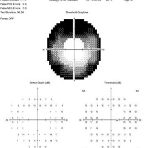 Birdshot Case # 2, Humphrey Visual Vield 60-4, OD
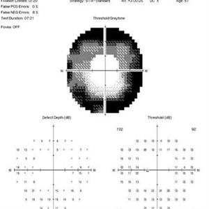
May 1 2013 by Armando L. Oliver, MD
57-year-old woman with known history of Birdshot Chorioretinopathy. Despite 20/20 best corrected visual acuity. The 60-4 Humphrey Visual Field reveals the extensive peripheral visual field loss characteristic of the condition.
Imaging device: Humphrey Visual Field
Condition/keywords: birdshot, birdshot chorioretinopathy, birdshot retinochoroidopathy
-
 Birdshot Case # 2, IVFA OD
May 1 2013 by Armando L. Oliver, MD
57-year-old Puerto Rican Woman presented for evaluation given history of chronic macular edema. Fundus examination revealed oval orange lesions dispersing from the disk. The patient has history of Type 2 DM. Work-up revealed that she was HLA-A29 positive. The patient had a negative CXR, FTA-Abs and RPR.
Photographer: Hector Mendez Caratini
Imaging device: Topcon TRC 50-DX
Condition/keywords: birdshot, birdshot chorioretinopathy, birdshot retinochoroidopathy
-
 Birdshot Case # 2, IVFA OS
May 1 2013 by Armando L. Oliver, MD
57-year-old Puerto Rican woman presented for evaluation given history of chronic macular edema. Fundus examination revealed oval orange lessions dispersing from the disk. The patient has history of Type 2 DM. Work-up revealed that she was HLA-A29 positive. The patient had a negative CXR, FTA-Abs and RPR.
Photographer: Hector Mendez Caratini
Imaging device: Topcon TRC 50DX
Condition/keywords: birdshot, birdshot chorioretinopathy, birdshot retinochoroidopathy
-
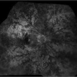
 Birdshot Case #1 OD FAF
May 1 2013 by Armando L. Oliver, MD
64-year-old Puerto Rican woman consulted due to the presence of 1+ vitreous cells. The fundus examination revealed orange to yellow lesions dispersing from the disk. Work-up revealed she was HLA-A29 positive and the suspected diagnosis of Birdshot Chorioretinopathy was made. Chest X-Ray, FTA-Abs and RPR were negative.
Photographer: Moises Castro, Instituto de Ojos y Piel, Carolina, PR
Imaging device: Zeiss, Visucam NM/FA
Condition/keywords: birdshot chorioretinopathy, birdshot retinochoroidopathy
-
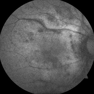
 Birdshot Case #1 OS FAF
May 1 2013 by Armando L. Oliver, MD
64-year-old Puerto Rican woman consulted due to the presence of 1+ vitreous cells. The fundus examination revealed orange to yellow lesions dispersing from the disk. Work-up revealed she was HLA-A29 positive and the suspected diagnosis of Birdshot Chorioretinopathy was made. Chest X-Ray, FTA-Abs and RPR were negative.
Photographer: Moises Castro, Instituto de Ojos y Piel, Carolina, PR
Imaging device: Zeiss, Visucam NM/FA
Condition/keywords: birdshot, birdshot chorioretinopathy, birdshot retinochoroidopathy
-
 Birdshot Case #2, HVF 60-4, OS
May 1 2013 by Armando L. Oliver, MD
57-year-old woman with known history of Birdshot Chorioretinopathy. Despite 20/20 best corrected visual acuity. The 60-4 Humphrey Visual Field reveals the extensive peripheral visual field loss characteristic of the condition.
Imaging device: Humphrey Visual Field 60-4
Condition/keywords: birdshot, birdshot chorioretinopathy, birdshot retinochoroidopathy
-
 Birdshot Chorioretinitis / Vitiliginous
Nov 7 2014 by David Callanan, MD
68-year-old female, birdshot chorioretinitis / vitiliginous.
Condition/keywords: birdshot chorioretinopathy, vitiliginous
-
 Birdshot Chorioretinopathy
Jul 26 2018 by Maxwell D Elia, MD
Fundus photo of 41-year-old HLA A29+ man with birdshot. He was referred after a period of 3 years of floaters and flashes.
Condition/keywords: birdshot chorioretinopathy
 Loading…