Search results (10 results)
-
 Achromatopsia / Rod monochromat
Apr 15 2014 by David Callanan, MD
1-year-old patient, achromatopsia / rod monochromat.
Condition/keywords: achromatopsia, rod monochromat
-
 Achromatopsia / Rod Monochromat
Apr 15 2014 by David Callanan, MD
1-year-old patient, achromatopsia / rod monochromat.
Condition/keywords: achromatopsia, rod monochromat
-
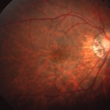
 Achromatopsia, Left Eye
Oct 28 2019 by Albert Li, MD, FASRS
Fundus photograph of 38-year-old man with congenital horizontal nystagmus.
Condition/keywords: achromatopsia
-
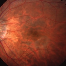
 Achromatopsia, right eye
Oct 28 2019 by Albert Li, MD, FASRS
Fundus photograph of 38-year-old man with congenital horizontal nystagmus.
Condition/keywords: achromatopsia
-
 Incomplete Achromatopsia
Mar 26 2019 by Gary R. Cook, MD, FACS
Right eye of a 15-year-old male with incomplete achromatopsia; V.A.= 20/20-3.
Imaging device: Topcon VT-50
Condition/keywords: achromatopsia
-
 Incomplete Achromatopsia
Mar 27 2019 by Gary R. Cook, MD, FACS
Left eye of a 15-year-old male with incomplete achromatopsia; V.A.= 20/50-2.
Imaging device: Topcon VT-50
Condition/keywords: achromatopsia
-
 OCT of Achromatopsia, Left Eye
Oct 28 2019 by Albert Li, MD, FASRS
OCT of 38-year-old man with congenital horizontal nystagmus with central ellipsoid zone loss.
Imaging device: Heidelberg Spectralis
Condition/keywords: achromatopsia
-
 OCT of Achromatopsia, Right Eye
Oct 28 2019 by Albert Li, MD, FASRS
OCT of 38-year-old man with congenital horizontal nystagmus with central ellipsoid zone loss.
Imaging device: Heidelberg Spectralis
Condition/keywords: achromatopsia
-
 OCT of Achromatopsia, Right Eye
Oct 28 2019 by Albert Li, MD, FASRS
OCT of 38-year-old man with congenital horizontal nystagmus with central ellipsoid zone loss.
Imaging device: Heidelberg Spectralis
Condition/keywords: achromatopsia
-
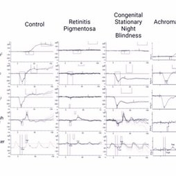 Representative Full Field Electroretinography Responses
May 13 2024 by Gabrielle Hallai
The left most column are control full field ERG responses from an individual with no known retinal pathology. In the second column is an example from a patient with autosomal recessive retinitis pigmentosa. This is an example of an intermediate case where rod function is extinguished but some cone function remains. In more advanced cases, full field ERG responses are typically extinguished to both scotopic and photopic stimuli. The third column is an example of congenital stationary night blindness (CSNB). While full field ERG responses can vary greatly depending on the specific subtype, this example of “complete CSNB” demonstrates extinguished rod pathway responses with the classic electronegative response for the scotopic 3.0 and 10.0 responses, consistent with bipolar cell dysfunction. Photopic cone responses are largely normal in this instance, but ”incomplete CSNB” can cause reduced photopic responses. In the final column, an example of full field ERG responses from a patient with achromatopsia. In achromatopsia, cone function is extinguished early in life, while rod pathway function is largely normal. ERG testing was completed using the Diagnosys ColorDome.
Photographer: Gabrielle Hallai, PhD, Cleveland Clinic Cole Eye Institute
Imaging device: Diagnosys ColorDome
Condition/keywords: achromatopsia, congenital stationary night blindness (CSNB), electroretinography, full field ERG, retinitis pigmentosa
 Loading…