Search results (402 results)
-
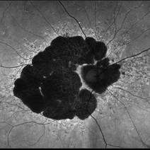
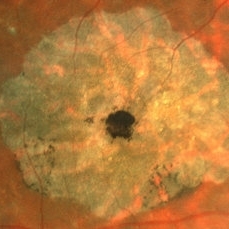
 B-FAF in Stargardt's Disease
Jul 4 2024 by Tejaswita Verma
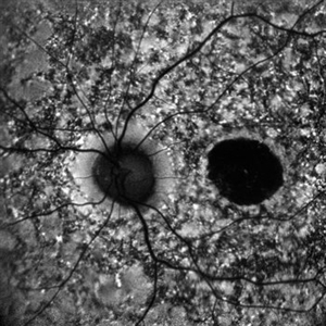
Blue fundus autofluorescence showing hypoautofluorescence picture of a 28 year old male with 6/60 vision in BE in a case of Stargardt's disease.
Photographer: DR. TEJASWITA VERMA
Imaging device: MIRANTE
Condition/keywords: fundus autofluorescence (FAF), hereditary macular dystrophy, Stargardt disease
-
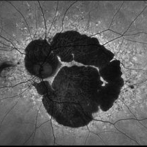
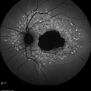
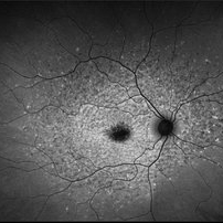
 Late Stage Stargardt's Disease
Mar 13 2013 by Hamid Ahmadieh, MD
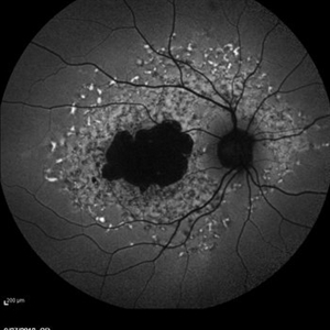
Autofluorescence imaging of the right eye of a 46-year-old man with decreased VA due to advanced Stargardt's disease.
Photographer: Nayereh Hadipoor, Negah Eye Center, Tehran
Imaging device: Heidelberg Spectralis
Condition/keywords: autofluorescence imaging, Stargardt disease
-
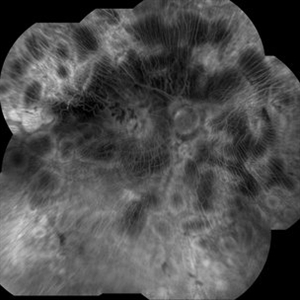
 Not All Stars Are in the Sky — Some Live in the Eyes of Those Learning to See in New Ways
Apr 21 2025 by rohan jain
Stargardt disease
Photographer: Dr. ROHAN JAIN
Condition/keywords: fleck retinopathy, fundus autofluorescence (FAF), hereditary macular dystrophy
-
 Stargardt Disease
Oct 7 2021 by Becca Harris
83-year-old male with Stargardt Disease.
Photographer: Becca Harris
Imaging device: Optos California
Condition/keywords: Optos, Stargardt disease
-
 Stargardt Disease
Oct 7 2021 by Becca Harris
83-year-old male with Stargardt Disease.
Photographer: Becca Harris
Imaging device: Optos California
Condition/keywords: Optos, Stargardt disease
-
 Stargardt Disease
Oct 7 2021 by Becca Harris
83-year-old male with Stargardt Disease.
Photographer: Becca Harris
Imaging device: Optos California
Condition/keywords: Optos, Stargardt disease
-
 Stargardt Disease
Oct 7 2021 by Becca Harris
83-year-old male with Stargardt Disease.
Photographer: Becca Harris
Imaging device: Optos California
Condition/keywords: left eye, Optos, Stargardt disease
-
 Stargardt Disease
Jan 20 2021 by Hannah Keller
Stargardt on 20-year-old male.
Photographer: Hannah Keller, Retina Specialists of Michigan
Imaging device: Optos
Condition/keywords: retina, Stargardt disease
-
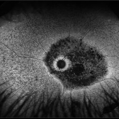
 Stargardt's disease
May 2 2013 by Henry J. Kaplan, MD
Typical Bull's eye.
Condition/keywords: bull's eye maculopathy, Stargardt disease
-
 Stargardt's Disease
Jun 11 2016 by John S. King, MD
Outer atrophy centrally with extrafoveal hyper-reflective protrusions into the outer retina.
Condition/keywords: bull's eye maculopathy, fleck dystrophy, Stargardt disease
-
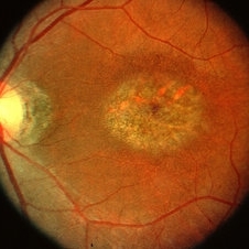
 Stargardt's Disease
Oct 4 2018 by Aditya S Kelkar, MS, FRCS, FASRS,FRCOphth
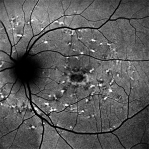
Auto-fluorescence image of a 19-year-old male showing flecks of both increased and decreased autofluorescence and reduced central macular autofluorescence surrounded by an increased signal.
Photographer: Dr. Aanchal Agarwal
Condition/keywords: Stargardt disease
-
 Stargardt's Disease
Oct 23 2024 by Virginia Gebhart
62 year old female with bullseye RPE changes and flecks, mottled FAF, and silent choroid on FA consistent with late onset Stargardt's Disease. Pt is asymptomatic with 20/20 vision OU at this time
Photographer: Virginia Gebhart, Retina Consultants of Carolina
Imaging device: Optos California
Condition/keywords: Stargardt disease, Stargardts Disease
-
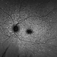
 Late Stage Stargardt's Disease
Mar 13 2013 by Hamid Ahmadieh, MD
Autofluorescence imaging of the left eye of a 46-year-old man with decreased VA due to advanced Stargardt's disease.
Photographer: Nayereh Hadipoor, Negah Eye Center, Tehran
Imaging device: Heidelberg Spectralis
Condition/keywords: autofluorescence imaging, Stargardt disease
-
 Stargardt macular dystrophy slide 2
Oct 22 2012 by Ronald C. Gentile, MD
The central areas of atrophy were symmetric in both eyes with increased visibility of the choroidal vessels within the atrophic macula.
Photographer: The New York Eye & Ear Infirmary Department of Medical Imaging
Condition/keywords: Stargardt disease
-
 Stargardt's Disease
Oct 13 2012 by Geoffrey G. Emerson, MD, PhD, FASRS
Stargardt's disease
Condition/keywords: Stargardt disease
-
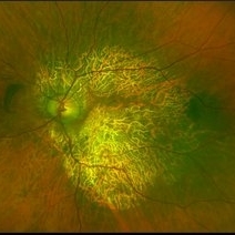
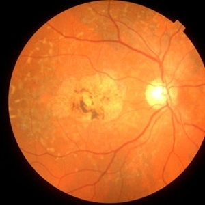 Late Stage Stargardt's Disease
Mar 13 2013 by Hamid Ahmadieh, MD
Color fundus photograph of the right eye of a 46-year-old man with decreased VA due to advanced Stargardt's disease.
Photographer: Nayereh Hadipoor, Negah Eye Center, Tehran
Imaging device: Heidelberg Spectralis
Condition/keywords: Stargardt disease
-
 Stargardt Disease
Feb 28 2013 by Theodore Leng, MD, MS, FASRS
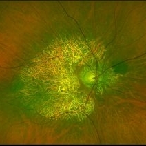
Fundus photograph of a 63-year-old man with Stargardt disease. Multiple pisciform flecks are visible.
Imaging device: Zeiss FF450
Condition/keywords: flecks, Stargardt disease
-
 Stargardt Disease
Feb 28 2013 by Theodore Leng, MD, MS, FASRS
Fundus photograph of a 63-year-old man with Stargardt disease. Multiple pisciform flecks are visible.
Imaging device: Zeiss FF450
Condition/keywords: flecks, Stargardt disease
-
 Stargardt's disease
May 2 2013 by Henry J. Kaplan, MD
The same patient , left eye #2
Condition/keywords: bull's eye maculopathy, Stargardt disease
-
 Stargardt's Disease
May 27 2020 by Jamin S. Brown, MD
Fundus autofluorescence image of 46-year-old male with Stargardt's Disease.
Photographer: Jeffrey Barker, Retina-Vitreous Surgeons of CNY
Condition/keywords: Stargardt disease
-
 Stargardt's Disease
Oct 18 2012 by Raj K. Maturi, MD
Photographer: Stephanie Morrow
Imaging device: HRA
Condition/keywords: Stargardt disease
-
 Stargardt's Disease
Oct 18 2012 by Raj K. Maturi, MD
Photographer: Stephanie Morrow
Imaging device: HRA
Condition/keywords: Stargardt disease
-
 Stargardt's Disease
Oct 18 2012 by Raj K. Maturi, MD
Photographer: Stephanie Morrow
Imaging device: HRA
Condition/keywords: red-free, Stargardt disease
-
 Stargardt’s Disease
Jul 21 2020 by William A. Townsend-Pico, MD
Fundus photograph and Fundus autofluoresence of a 21-year-old female with progressive loss of vision OU.
Photographer: Dania Otero, Retina Consultants of Puerto Rico
Imaging device: Optos California Fundus Camera
Condition/keywords: Stargardt disease
-
 Stargardt’s Disease
Jul 21 2020 by William A. Townsend-Pico, MD
Fundus photograph and Fundus autofluoresence of a 21-year-old female with progressive loss of vision OU.
Photographer: Dania Otero, Retina Consultants of Puerto Rico
Imaging device: Optos California Fundus Camera
Condition/keywords: Stargardt disease
 Loading…