Search results (28 results)
-
 Giant RPE-rip
Sep 5 2021 by Hemanth Murthy, MBBS, MD, FASRS
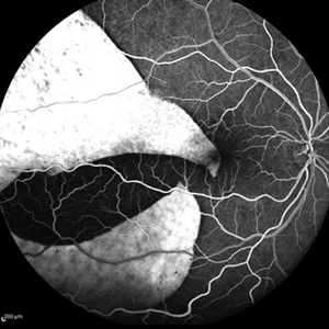
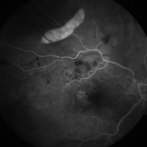
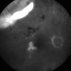
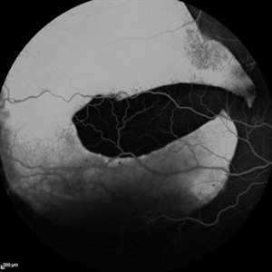
Fundus fluorescein angiography of a 50 year-old patient with spontaneous giant RPE rip.
Photographer: Mr Veda Vyas
Imaging device: Heidelberg HRA
Condition/keywords: RPE-Rip
-
 Extrafoveal PED with RPE rip FA3
Dec 23 2012 by Alex P. Hunyor, MD
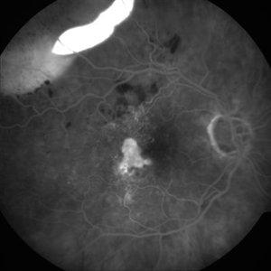
80-year-old female with subfoveal occult CNV and large extrafoveal PED which underwent spontaneous RPE rip. FA shows intense hyperfluorescence in area of absent RPE, progressive filling of extrafoveal PED, and hyperfluorescence in macula from atrophy and occult CNV.
Condition/keywords: pigment epithelial detachment (PED), retinal pigment epithelium (RPE) tear
-
 ARMD with RPE Rip
Oct 12 2012 by Jeffrey G. Gross, MD, FASRS
ARMD with RPE rip.
Condition/keywords: retinal pigment epithelium, retinal pigment epithelium (RPE) tear
-
 ARMD with RPE Rip
Oct 12 2012 by Jeffrey G. Gross, MD, FASRS
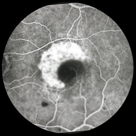
ARMD with RPE rip, FA, showing window defect and blockage from retracted RPE layer.
Condition/keywords: retinal pigment epithelium, retinal pigment epithelium (RPE) tear, retracted retinal pigment epithelium (RPE) layer
-
 Extrafoveal PED with RPE rip AF
Dec 23 2012 by Alex P. Hunyor, MD
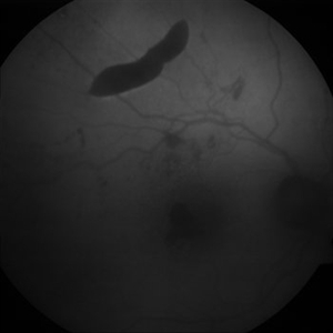
80-year-old female with subfoveal occult CNV and large extrafoveal PED which underwent spontaneous RPE rip. Autofluorescence image shows hypoautofluorescence in crescentic area of absent RPE due to rip, and also RPE atrophy adjacent to fovea. Intervening small areas of hypoautofluorescence are due to subretinal haemorrhage.
Condition/keywords: pigment epithelial detachment (PED), retinal pigment epithelium (RPE) tear
-
 Extrafoveal PED with RPE rip colour photo
Dec 23 2012 by Alex P. Hunyor, MD
80-year-old female with subfoveal occult CNV and large extrafoveal PED which underwent spontaneous RPE rip.
Condition/keywords: pigment epithelial detachment (PED), retinal pigment epithelium (RPE) tear
-
 Extrafoveal PED with RPE rip FA1
Dec 23 2012 by Alex P. Hunyor, MD
80-year-old female with subfoveal occult CNV and large extrafoveal PED which underwent spontaneous RPE rip. Early phase FA showing intense hyperfluorescence in the area of acute absence of RPE.
Condition/keywords: pigment epithelial detachment (PED), retinal pigment epithelium (RPE) tear
-
 Extrafoveal PED with RPE rip FA2
Dec 23 2012 by Alex P. Hunyor, MD
80-year-old female with subfoveal occult CNV and large extrafoveal PED which underwent spontaneous RPE rip. FA shows intense hyperfluorescence in area of absent RPE, progressive filling of extrafoveal PED, and hyperfluorescence in macula from atrophy and occult CNV.
Condition/keywords: pigment epithelial detachment (PED), retinal pigment epithelium (RPE) tear
-
 Extrafoveal PED with RPE rip FA4
Dec 23 2012 by Alex P. Hunyor, MD
80-year-old female with subfoveal occult CNV and large extrafoveal PED which underwent spontaneous RPE rip. FA shows intense hyperfluorescence in area of absent RPE, progressive filling of extrafoveal PED, and hyperfluorescence in macula from atrophy and occult CNV.
Condition/keywords: pigment epithelial detachment (PED), retinal pigment epithelium (RPE) tear
-
 Extrafoveal PED with RPE rip FA5
Dec 23 2012 by Alex P. Hunyor, MD
80-year-old female with subfoveal occult CNV and large extrafoveal PED which underwent spontaneous RPE rip. FA shows intense hyperfluorescence in area of absent RPE, progressive filling of extrafoveal PED, and hyperfluorescence in macula from atrophy and occult CNV.
Condition/keywords: pigment epithelial detachment (PED), retinal pigment epithelium (RPE) tear
-
 Fluorescein Angiogram of an RPE Rip
Feb 21 2022 by Maxwell J Wingelaar, MD
A 88-year-old male with a history of AMD who presented with an RPE rip
Photographer: Jarrod Wehmeier
Condition/keywords: RPE Rip
-
 Giant RPE -Rip
Sep 5 2021 by Hemanth Murthy, MBBS, MD, FASRS
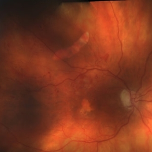
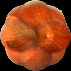
Fundus photo OD of 50 year-old patient with spontaneous giant RPE rip.
Photographer: Mr Veda Vyas
Imaging device: Heidelberg HRA
Condition/keywords: Giant RPE-Rip, RPE-Rip
-
 Giant RPE rip
Sep 5 2021 by Hemanth Murthy, MBBS, MD, FASRS
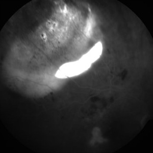
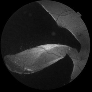
Autofluorescence of a 50 year-old patient with spontaneous giant RPE rip.
Photographer: Mr Veda Vyas
Imaging device: Heidelberg HRA
Condition/keywords: RPE-rip
-
 Giant RPE-Rip
Sep 5 2021 by Hemanth Murthy, MBBS, MD, FASRS
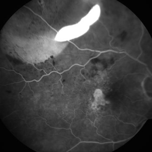
Fundus fluorescein angiography of a 50 year-old patient with spontaneous giant RPE rip.
Photographer: Mr Veda Vyas
Imaging device: Heidelberg HRA
Condition/keywords: RPE-Rip
-
 RPE RIP
Apr 2 2019 by Gary R. Cook, MD, FACS
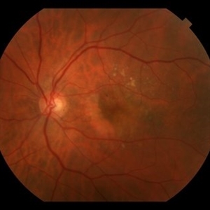
Elderly white female with exudative AMD and RPED with a RPE RIP OS; VA = 20/80.
Condition/keywords: retinal pigment epithelium, retinal pigment epithelium (RPE) contracture
-
 RPE RIP
Jun 6 2019 by Gary R. Cook, MD, FACS
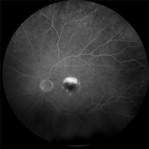
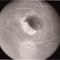
Mid-phase fluorescein angiogram of an elderly white female with exudative AMD and a RPE rip OS; V.A. = 20/80
Condition/keywords: FA mid phase, fluorescein angiogram (FA), retinal pigment epithelium
-
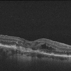
 RPE Rip
Aug 21 2024 by Amirfarbod Yazdanyar, MD , PhD
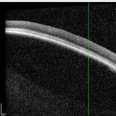
OCT image of PED with RPE-rip in 85 year-old male with history of AMD.
Condition/keywords: pigment epithelial detachment (PED), RPE-rip
-
 RPE Rip
Feb 13 2013 by From the Collections of Thomas M. Aaberg, MD and Thomas M. Aaberg Jr., MD
RPE rip, foveal CNV
Condition/keywords: choroidal neovascularization (CNV), macular laser, retinal pigment epithelium (RPE) tear
-
 RPE Rip
Jan 25 2024 by Virginia Gebhart
69 year old female with Neovascular AMD. New RPE rip and increased IRF on OCT 10 weeks s/p Eylea injection. Switched to Vabysmo to extend intervals
Photographer: Virginia Gebhart
Imaging device: Topcon
Condition/keywords: neovascular age-related macular degeneration (AMD)
-
 RPE rip in a case of Idiopathic polypoidal choroidopathy
Oct 23 2022 by Anjana Mirajkar, MS Ophthalmology
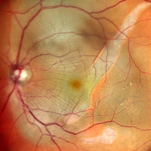
Color photo central image in a of 61 year old male with RPE rip in a case of Idiopathic Polypoidal Choroidopathy.
Photographer: Dr. Anjana Mirajkar -Retina Foundation, Ahmedabad
Condition/keywords: idiopathic polypoidal choroidopathy, RPE Rip
-
 RPE rip in a case of Idiopathic polypoidal choroidopathy
Oct 23 2022 by Anjana Mirajkar, MS Ophthalmology
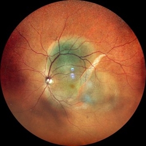
Color photo wide field image in a of 61 year old male with RPE rip in a case of Idiopathic Polypoidal Choroidopathy.
Photographer: Dr. Anjana Mirajkar -Retina Foundation, Ahmedabad
Condition/keywords: RPE Rip
-
 RPE rip in a case of Idiopathic polypoidal choroidopathy
Oct 23 2022 by Anjana Mirajkar, MS Ophthalmology
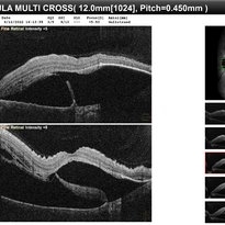
OCT image in a of 61 year old male with RPE rip in a case of Idiopathic Polypoidal Choroidopathy.
Photographer: Dr. Anjana Mirajkar -Retina Foundation, Ahmedabad
Condition/keywords: Idiopathic polypoidal choroidopathy, RPE rip
-
 RPE rip in a case of Idiopathic polypoidal choroidopathy
Oct 23 2022 by Anjana Mirajkar, MS Ophthalmology
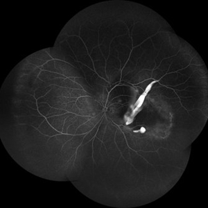
Montage of Fluorescein angiography in a of 61 year old male with RPE rip in a case of Idiopathic Polypoidal Choroidopathy.
Photographer: Dr. Anjana Mirajkar -Retina Foundation, Ahmedabad
Condition/keywords: Idiopathic polypoidal choroidopathy, RPE rip
-
 RPE rip macular OCT
Dec 23 2012 by Alex P. Hunyor, MD
80-year-old female with subfoveal occult CNV and large extrafoveal PED which underwent spontaneous RPE rip. OCT shows subfoveal CNV and intraretinal cystic edema
Condition/keywords: pigment epithelial detachment (PED), retinal pigment epithelium (RPE) tear
-
---thumb.jpg/image-square;max$300,300.ImageHandler) Subretinal Neovascular Membrane With RPE
Subretinal Neovascular Membrane With RPE
Oct 11 2013 by Maurice F. Rabb
This healthy 81 year old Caucasian female has a family history of macular degeneration. She has no history of hypertension, and has not been on ASA or anticoagulants. She had a subretinal neovascular membrane with an RPE rip of the left eye, which advanced to disciform macular degeneration with eventual massive hemorrhage and total exudative retinal detachment. She developed neovascular glaucoma OS due to total retinal detachment and had enucleation of the left eye.
Condition/keywords: subretinal neovascular membrane
 Loading…