Search results (21 results)
-
 Retinal Detachment with PVR (s/ SPR, PPV, MPV, 360 Retinectomy, PFO, PI, FAx, SO)
Aug 22 2019 by Merrick Avila
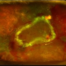
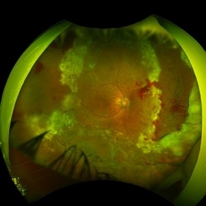
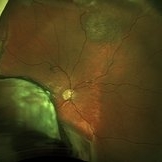
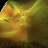
Ultra-wide field pseudocolor fundus photograph of a 64-year-old female with a treated retinal detachment with proliferative vitreoretinopathy. Patient has a history of complex retinal detachments that have been treated multiple times. On exam 8-22-19, there were large macular holes with LP vision. There was a long discussion about guarded nature of her condition and goals or trial for repair including globe sparing prevention of phthisis.
Photographer: Merrick Avila
Imaging device: Optos
Condition/keywords: diabetic retinopathy, hemorrhage, Optos, proliferative vitreoretinopathy (PVR), retinectomy, silicone oil
-
 Submacular PFO
Feb 20 2020 by Kevin J. Blinder, MD, FASRS
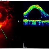
This is a 53-year-old gentleman that was referred to us for a second opinion with an inoperable RD with PVR after 3 failed attempts. We performed a PPV, membranectomy, scleral buckling procedure, with silicone oil injection. This case did not require PFO. You can imagine our surprise when we discovered submacular PFO postoperatively. It is very difficult to see the PFO on the Optos. The infrared shows it clearly, with confirmation of the submacular space on the SD-OCT.
Photographer: Jarrod Wehmeier, The Retina Institute; St. Louis, MO
Imaging device: optos
Condition/keywords: submacular perfluorocarbon liquid (PFO)
-
 PFO Bubbles
Feb 25 2025 by Parnian Arjmand, MD, MSc, FRCSC, DABO
Post operative day 7 after repair of an RD secondary to a giant retinal tear with temporary PFO tamponade.
Condition/keywords: GRT, PFO
-
 submacular perfluorocarbon liquid
Sep 7 2022 by JEFFERSON R SOUSA, Tecg.º (Biomedical Systems Technology)
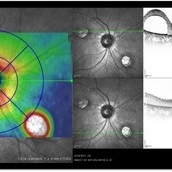
A 63-year-old male patient underwent vitreoretinal surgery with the use of perfluorocarbon. From a technological point of view, extended-field retinography presents many points of focus variation due to the difficulty of establishing a diffuse focus, as it is a recent post-operative case. In OCT Fundus Enface, although it has a low resolution, it is extremely important for documenting the presence of perfluor. Best seen in structural OCT.
Photographer: JEFFERSON ROCHA DE SOUSA - Retinal Department at Instituto Dr. Suel Abujamra Sao Paulo-Brazil
Imaging device: Optical Coherence Tomography system OCT CIRRUS 5000, Protocol, HD 5 Line
Condition/keywords: perfluorocarbon fluid, post-vitrectomy, submacular perfluorocarbon liquid (PFO), vitrectomy
-
 ILM Removal
Apr 5 2018 by Mohamed Tawfik, MD
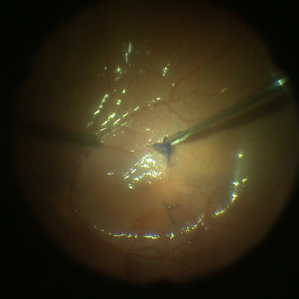
Steps Of ILM peel stained with brilliant blue under PFO.
Photographer: Mohamed A,Tawfik MD,FRSCed
Imaging device: intra opeative Photography Screen shoot
Condition/keywords: internal limiting membrane (ILM) peeling
-
 Peeling Under PFO
May 7 2023 by Maxwell J Wingelaar, MD
Peeling under PFO
Condition/keywords: ILM peeling, ILM staining, proliferative vitreoretinopathy (PVR)
-
 Retinal Detachment Following Scleral Buckling, Retinectomy, Laser, and Oil
Jan 31 2022 by Ahmad B. Tarabishy, MD
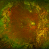
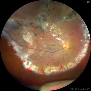
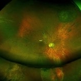
Ultra wide-field fundus photograph of a 55-year-old gentleman who is 4 days after surgery with scleral buckling, pars plana vitrectomy, perfluoron tamponade, membrane peeling, direct fluid-PFO-oil exchange, nasal and temporal retinectomies, and endolaser photocoagulation. Visual acuity was 20/150 under oil.
Photographer: Megan McLandsborough, Lakeland Eye Clinic
Imaging device: Optos California UWF Camera
Condition/keywords: endolaser, Membrane Peel, PPV, proliferative retinopathy, proliferative vitreoretinopathy (PVR), Retinal Detachment, retinal detachment with retinal defect, scleral buckle, submacular perfluorocarbon liquid (PFO)
-
 submacular perfluorocarbon liquid
Sep 7 2022 by JEFFERSON R SOUSA, Tecg.º (Biomedical Systems Technology)
A 63-year-old male patient underwent vitreoretinal surgery with the use of perfluorocarbon. From a technological point of view, extended-field retinography presents many points of focus variation due to the difficulty of establishing a diffuse focus, as it is a recent post-operative case. In OCT Fundus Enface, although it has a low resolution, it is extremely important for documenting the presence of perfluor. Best seen in structural OCT.
Photographer: JEFFERSON ROCHA DE SOUSA - Retinal Department at Instituto Dr. Suel Abujamra Sao Paulo-Brazil
Imaging device: Clarus 700 - Zeiss, 135 degree images.
Condition/keywords: perfluorocarbon fluid, post-vitrectomy, submacular perfluorocarbon liquid (PFO), vitrectomy
-
 Submacular PFO
Feb 20 2020 by Kevin J. Blinder, MD, FASRS
This is a 53-year-old gentleman that was referred to us for a second opinion with an inoperable RD with PVR after 3 failed attempts. We performed a PPV, membranectomy, scleral buckling procedure, with silicone oil injection. This case did not require PFO. You can imagine our surprise when we discovered submacular PFO postoperatively. It is very difficult to see the PFO on the Optos. The infrared shows it clearly, with confirmation of the submacular space on the SD-OCT.
Photographer: Jarrod Wehmeier, The Retina Institute; St. Louis, MO
Imaging device: Heidelberg Spectralis
Condition/keywords: submacular perfluorocarbon liquid (PFO)
-
 24 Hours Post Scleral Wound Closure+ Scleral Buckle+25 g Vitrectomy+Silicon Oil
Jan 23 2015 by Carlos Quezada-Ruiz, MD, FASRS
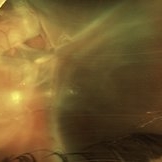
24 hours post op fundus photograph of a 43-year-old man who had perforating injury to the right eye with a small piece of plastic while he was hammering. OD LP, subconjunctival hemorrhage, clear cornea, hyphema, irido and ciclodyalisis as well as a luxated lens with traumatic cataract and a dense vitreous hemorrhage. B-US showed rhegmatogenous retinal detachment with a tear and a big inferior hemorrhagic choroidal detachment. 360 peritomy revealed 2-entry scleral wounds were found in zone II (M V and M VI) and closure was performed. 25 G PPV was performed with the infusion canal placed in the AC through the limbus. Lensectomy and removal of a dense recent vitreous hemorrhage revealed a white detached retina with an exit wound through the temporal inferior segment of the optic nerve with a nasal GRT and sub retinal hemorrhage as well as temporal inferior choroidal, PVD was induced and PFOs helped stabilizing the retina while vitrectomy and sub-retinal hemorrhage was removed through the GRT. Fluid air exchange was made and 360 endolaser over the buckle indentation was done and silicon oil was used as endotamponade. This picture was taken 24 hrs after the surgery.
Photographer: Lilibeth Rodriguez, Instituto de la Visión. Torreon, Mexico.
Condition/keywords: central retinal artery occlusion (CRAO), giant retinal tear, trauma
-
 blue2
blue2
Jun 30 2012 by Stanislao Rizzo, MD
PFO associated with blue staining facilitates the PVR membranes removal
-
 PFO Bubbles
Oct 24 2024 by Korey Starkey
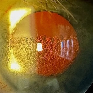
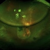
Slit lamp photograph of a 23 year old female with PFO bubbles inferiorly in the AC. Discussed surgical intervention to remove PFO from AC and vitreous cavity in future.
Photographer: Korey Starkey
Imaging device: Slit lamp camera
Condition/keywords: anterior chamber, PFO, slit lamp photography, submacular perfluorocarbon liquid (PFO)
-
 POD 1
POD 1
Jan 7 2018 by John S. King, MD
61-year-old developed a large choroidal OD after a bleb revision. 30 years ago had an IOFB removed. The initial photos, POD 1 and POW 3 photos are provided. Dr. Zocchi performed the surgery. Surgery included 42 band placed 360 (superior bleb was fibrosed and left in place during conj peritomy); suprachoroidal drainiage was via radial incision; a pre-existing ST tear was present; the retinal adhesions were removed with blunt dissection, then remaining pvr was peeled; PFO used to flatten retina; 5000 cs sil oil used. Post-op vision has improved for near to J5.
Imaging device: Optos
Condition/keywords: suprachoroidal hemorrhage
-
 POW 3
POW 3
Jan 7 2018 by John S. King, MD
61-year-old developed a large choroidal OD after a bleb revision. 30 years ago had an IOFB removed. The initial photos, POD 1 and POW 3 photos are provided. Dr. Zocchi performed the surgery. Surgery included 42 band placed 360 (superior bleb was fibrosed and left in place during conj peritomy); suprachoroidal drainiage was via radial incision; a pre-existing ST tear was present; the retinal adhesions were removed with blunt dissection, then remaining pvr was peeled; PFO used to flatten retina; 5000 cs sil oil used. Post-op vision has improved for near to J5.
Imaging device: Optos
Condition/keywords: suprachoroidal hemorrhage
-
 Pre-OP
Jan 7 2018 by John S. King, MD
61-year-old developed a large choroidal OD after a bleb revision. 30 years ago had an IOFB removed. The initial photos, POD 1 and POW 3 photos are provided. Dr. Zocchi performed the surgery. Surgery included 42 band placed 360 (superior bleb was fibrosed and left in place during conj peritomy); suprachoroidal drainiage was via radial incision; a pre-existing ST tear was present; the retinal adhesions were removed with blunt dissection, then remaining pvr was peeled; PFO used to flatten retina; 5000 cs sil oil used. Post-op vision has improved for near to J5.
Imaging device: Optos
Condition/keywords: suprachoroidal hemorrhage
-
 Sub macular perfluorocarbon liquid
Mar 6 2022 by mohamed ahmed el shafie, ophthalmic resident
postoperative Sub macular perfluorocarbon liquid
Condition/keywords: submacular perfluorocarbon liquid (PFO)
-
 submacular perfluorocarbon liquid
Sep 7 2022 by JEFFERSON R SOUSA, Tecg.º (Biomedical Systems Technology)
A 63-year-old male patient underwent vitreoretinal surgery with the use of perfluorocarbon. From a technological point of view, extended-field retinography presents many points of focus variation due to the difficulty of establishing a diffuse focus, as it is a recent post-operative case. In OCT Fundus Enface, although it has a low resolution, it is extremely important for documenting the presence of perfluor. Best seen in structural OCT.
Photographer: JEFFERSON ROCHA DE SOUSA - Retinal Department at Instituto Dr. Suel Abujamra Sao Paulo-Brazil
Imaging device: Optical Coherence Tomography system OCT CIRRUS 5000, Protocol OCT Fundus Enface.
Condition/keywords: perfluorocarbon fluid, post-vitrectomy, submacular perfluorocarbon liquid (PFO), vitrectomy
-
 Submacular Perfluorocarbon Liquid Bubble
Apr 28 2021 by Marco Antonio Sauza
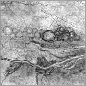
Red-free photo of a 70-year-old male with retinal detachment with a submacular perfluorocarbon liquid bubble after vitrectomy surgery.
Photographer: DR. MARCO SAUZA
Imaging device: ZEISS
Condition/keywords: submacular perfluorocarbon liquid (PFO)
-
 Subretinal PFO
Feb 7 2017 by Andrea Arriola-Lopez, MD MSc
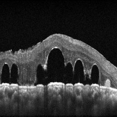
SD OCT of 32-year-old woman two weeks after vitrectomy. Round SRF was found. VA 20/50.
Photographer: Andrea Elizabeth Arriola-Lopez MD MSc
Imaging device: Heidelberg SD-OCT
Condition/keywords: perfluorocarbon fluid
 Loading…