Search results (309 results)
-
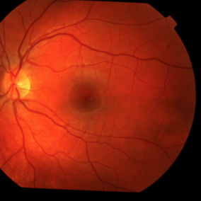
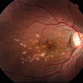 Multiple evanescent White Dot Syndrome (MEWDS)
May 27 2025 by César Adrián Gómez Valdivia, MD
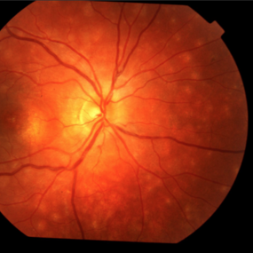
Fundus photograph of a 21 year-old female patient with suspected Multiple Evanescent White Dot Syndrome (MEWDS). The White Dot Syndromes produce yellow-white retinal lesions classically located at the retinal pigment epithelium or outer retina and are found primarily in young adults. Symptoms of MEWDS include unilateral blurred vision, visual field loss, photopsias, and floaters.
Photographer: @eyemissu2
Imaging device: TOPCON TRC-50DX
Condition/keywords: multiple evanescent white dot syndrome (MEWDS)
-
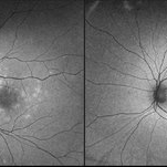 Resolved Multiple Evanescent White Dot Syndrome
Feb 18 2025 by Jordyn Beckman
Autofluorescence fundus photographs of Resolved Multiple Evanescent White Dot Syndrome in a 28 year old female with previous hyperfluorescent punctate spots throughout the posterior pole.
Photographer: Jordyn Beckman, Retina Consultants of Carolina, P.A.
Imaging device: California Optos
Condition/keywords: autofluorescence imaging, grey-white lesions, multiple evanescent white dot syndrome (MEWDS), scattered punctate
-
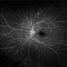 MEWDS
MEWDS
Dec 11 2024 by Virginia Gebhart
28 year old female with new Multiple Evanescent White Dot Syndrome. Patient reports gray spot in vision, OCT shows RPE disruption centrally but no edema. FA shows early hyperfluorescent punctate spots throughout the posterior pole, but no leakage. Normal findings OD. Will observe for now
Photographer: Virginia Gebhart, Retina Consultants of Carolina
Imaging device: Optos California
Condition/keywords: FA, fluorescein angiogram (FA), multiple evanescent white dot syndrome (MEWDS)
-
 Thrombocytopenia
Sep 24 2024 by DR Rohit Gupta
Fundus photography of a 16 year-old girl suffering from severe thrombocytopenia, showing flame shaped hemorrhage.
Photographer: Dr Rohit gupta
Imaging device: Samsung S21
Condition/keywords: anaemic retinopathy, flame shaped retinal hemorrhage, Haemorrhage, Roth spots, white centered retinal hemorrhage (Roth Spot), white dot syndrome
-
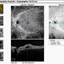 Acute Posterior Multifocal Placoid Pigment Epitheliopathy
Feb 20 2024 by Soobien Lee
12x12mm OCT Angiography of a 20-year-old caucasian female with viral prodrome and vision loss OS>OD secondary to Acute Posterior Multifocal Placoid Pigment Epitheliopathy (APPME). Imaging shows multifocal flow voids.
Photographer: Kim Seay, Elman Retina Group
Imaging device: 12x12mm OCT-Angiography
Condition/keywords: acute posterior multifocal placoid pigment epitheliopathy (APMPPE), bacillary layer detachment, OCT, OCT Angiography, Uveitis, white dot syndrome
-
 Acute Posterior Multifocal Placoid Pigment Epitheliopathy
Feb 20 2024 by Soobien Lee
A 20-year-old caucasian female with viral prodrome and vision loss OS>OD secondary to Acute Posterior Multifocal Placoid Pigment Epitheliopathy (APPME). OCT of the left macula shows bacillary layer detachment.
Photographer: Kim Seay, Elman Retina Group
Condition/keywords: acute posterior multifocal placoid pigment epitheliopathy (APMPPE), bacilliary layer detachment, OCT, Uveitis, white dot syndrome
-
 Acute Posterior Multifocal Placoid Pigment Epitheliopathy
Feb 20 2024 by Soobien Lee
Fluorescein angiogram of a 20-year-old caucasian female with viral prodrome and vision loss OS>OD secondary to Acute Posterior Multifocal Placoid Pigment Epitheliopathy (APPME). Early blockage with late hyperfluorescent leakage can be seen on fluorescein angiography of the left eye.
Photographer: Ashley Metzger, Elman Retina Group
Imaging device: Optos Ultra-Widefield Fluorescein Angiography
Condition/keywords: acute posterior multifocal placoid pigment epitheliopathy (APMPPE), bacilliary layer detachment, FA, FA late phase, FA late phase leakage, fluorescein angiogram (FA), Optos, uveitis, white dot syndrome
-
 Acute Posterior Multifocal Placoid Pigment Epitheliopathy
Feb 20 2024 by Soobien Lee
Fluorescein angiogram of a 20-year-old caucasian female with viral prodrome and vision loss OS>OD secondary to Acute Posterior Multifocal Placoid Pigment Epitheliopathy (APPME). Early blockage with late hyperfluorescent leakage can be seen on fluorescein angiography of the left eye.
Photographer: Ashley Metzger, Elman Retina Group
Imaging device: Optos Ultra-Widefield Fluorescein Angiography
Condition/keywords: acute posterior multifocal placoid pigment epitheliopathy (APMPPE), bacilliary layer detachment, FA, FA early phase, fluorescein angiogram (FA), Optos, uveitis, white dot syndrome
-
 Acute Posterior Multifocal Placoid Pigment Epitheliopathy
Feb 20 2024 by Soobien Lee
Optos fundus autofluorescence photograph of a 20-year-old caucasian female with viral prodrome and vision loss OS>OD secondary to Acute Posterior Multifocal Placoid Pigment Epitheliopathy (APPME). Imaging of her left eye shows hypoautofluorescent areas corresponding to multiple bilateral placoid lesions at the level of RPE and choroid throughout the posterior pole.
Photographer: Ashley Metzger, Elman Retina Group
Imaging device: Optos Ultra-Widefield Autoflurescence Imaging
Condition/keywords: acute posterior multifocal placoid pigment epitheliopathy (APMPPE), autofluorescence imaging, bacilliary layer detachment, Optos, OPTOS CALIFORNIA, uveitis, white dot syndrome
-
 Acute Posterior Multifocal Placoid Pigment Epitheliopathy
Feb 20 2024 by Soobien Lee
Optos color fundus photograph of a 20-year-old caucasian female with viral prodrome and vision loss OS>OD secondary to Acute Posterior Multifocal Placoid Pigment Epitheliopathy (APPME). Imaging of her left eye shows multiple bilateral creamy yellow-white placoid lesions at the level of RPE and choroid throughout the posterior pole.
Photographer: Ashley Metzger, Elman Retina Group
Imaging device: Optos Ultra-Widefield Imaging
Condition/keywords: acute posterior multifocal placoid pigment epitheliopathy (APMPPE), bacilliary layer detachment, Optos, uveitis, white dot syndrome
-
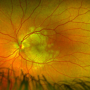
 MEWDs
MEWDs
Feb 28 2022 by Sruthi Arepalli, MD
Fundus photograph of a 37 year old woman presenting with photopsias and ellipsoid zone disruptions on OCT with findings consistent with MEWDS on autofluorescence. These areas of autofluorescence improved with oral steroids.
Condition/keywords: Autoflourescence, multiple evanescent white dot syndrome (MEWDS)
-
 Multiple Evanescent White Dot Syndrome
Feb 10 2021 by Cláudia Farinha
Ultra-widefield color and autofluorescence of a 30-year-old myopic female with decreased visual acuity, photopsias, and temporal scotomata.
Photographer: Claudia Farinha, MD
Imaging device: Optomap, Optos
Condition/keywords: multiple evanescent white dot syndrome (MEWDS)
-
 MEWDS
MEWDS
Oct 9 2020 by David L Kilpatrick, MD
26-year-old female presented with unilateral vision loss. She c/o flashes and a peripheral scotoma. Vision was 20/100. On exam, she showed foveal granularity, mild disc edema, and white dots as seen. Three weeks later, white dots had resolved and vision improved to 20/25.
Photographer: Mississippi Retina Associates
Imaging device: Optos
Condition/keywords: multiple evanescent white dot syndrome (MEWDS)
-
 MEWDS - Widefield FA
Oct 9 2020 by David L Kilpatrick, MD
26-year-old female presented with unilateral vision loss. She c/o flashes and a peripheral scotoma. Vision was 20/100. On exam, she showed foveal granularity, mild disc edema, and white dots as seen. Three weeks later, white dots had resolved and vision improved to 20/25.
Photographer: Mississippi Retina Associates
Imaging device: Optos
Condition/keywords: multiple evanescent white dot syndrome (MEWDS)
-
 Birdshot: a View From the Outside
Nov 3 2019 by Julia Farah, MD
61-year-old female presented with classic birdshot chorioretinopathy.
Photographer: Peter Guingab
Imaging device: Optos California
Condition/keywords: birdshot choroidopathy, uveitis, white dot syndrome
-
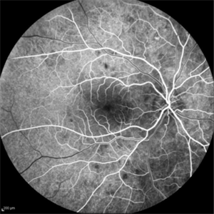 Multiple Evanescent White Dot Syndrome
Nov 1 2019 by Thomas A. Ciulla, MD, MBA, FASRS
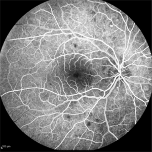
Fluorescein angiogram of an 18-year-old woman with multiple evanescent white dot syndrome (MEWDS). She noted a 5 day history of a new blind spot OD.
Condition/keywords: multiple evanescent white dot syndrome (MEWDS)
-
 Multiple Evanescent White Dot Syndrome
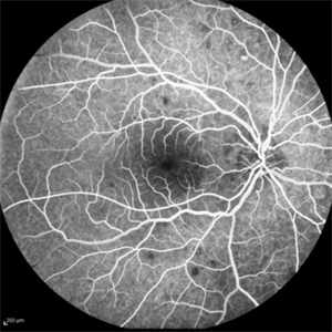
Nov 1 2019 by Thomas A. Ciulla, MD, MBA, FASRS
Fluorescein angiogram of an 18-year-old woman with multiple evanescent white dot syndrome (MEWDS). She noted a 5 day history of a new blind spot OD.
Condition/keywords: multiple evanescent white dot syndrome (MEWDS)
-
 Multiple Evanescent White Dot Syndrome
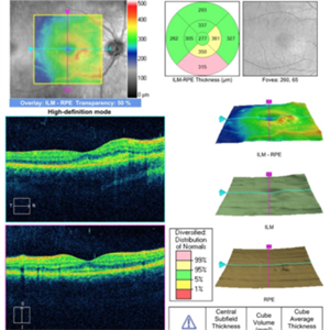
Nov 1 2019 by Thomas A. Ciulla, MD, MBA, FASRS
OCT OD of an 18-year-old woman with multiple evanescent white dot syndrome (MEWDS). She noted a 5 day history of a new blind spot OD.
Condition/keywords: multiple evanescent white dot syndrome (MEWDS)
-
 Multiple Evanescent White Dot Syndrome
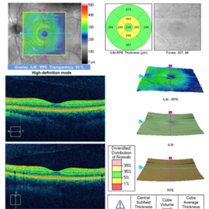
Nov 1 2019 by Thomas A. Ciulla, MD, MBA, FASRS
OCT OS of an 18-year-old woman with multiple evanescent white dot syndrome (MEWDS). She noted a 5 day history of a new blind spot OD.
Condition/keywords: multiple evanescent white dot syndrome (MEWDS)
-
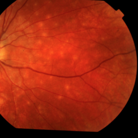
 Multiple Evanescent White Dot Syndrome (MEWDS)
Nov 1 2019 by Thomas A. Ciulla, MD, MBA, FASRS
Fundus photograph of an 18-year-old woman with multiple evanescent white dot syndrome (MEWDS). She noted a 5 day history of a new blind spot OD.
Condition/keywords: multiple evanescent white dot syndrome (MEWDS)
-
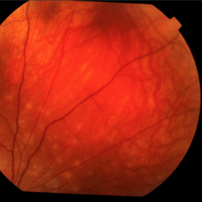
 Multiple Evanescent White Dot Syndrome (MEWDS)
Nov 1 2019 by Thomas A. Ciulla, MD, MBA, FASRS
Fundus photograph of an 18-year-old woman with multiple evanescent white dot syndrome (MEWDS). She noted a 5 day history of a new blind spot OD.
Condition/keywords: multiple evanescent white dot syndrome (MEWDS)
-
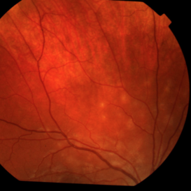
 Multiple Evanescent White Dot Syndrome (MEWDS)
Nov 1 2019 by Thomas A. Ciulla, MD, MBA, FASRS
Fundus photograph of an 18-year-old woman with multiple evanescent white dot syndrome (MEWDS). She noted a 5 day history of a new blind spot OD.
Condition/keywords: multiple evanescent white dot syndrome (MEWDS)
-
 Multiple Evanescent White Dot Syndrome (MEWDS)
Nov 1 2019 by Thomas A. Ciulla, MD, MBA, FASRS
Fundus photograph of an 18-year-old woman with multiple evanescent white dot syndrome (MEWDS). She noted a 5 day history of a new blind spot OD.
Condition/keywords: multiple evanescent white dot syndrome (MEWDS)
 Loading…