Search results (253 results)
-
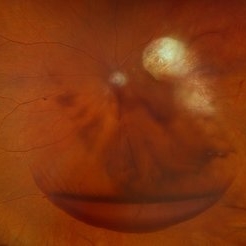
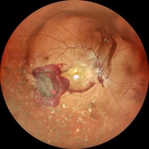 Subhyaloid Hemorrhage With Dispersed Vitreous Hemorrhage in a Case of Old Lasered Branch Retinal Vein Occlusion
Jul 12 2025 by Akansha Sharma
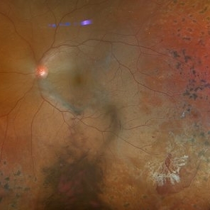
Color fundus photograph of a 32 year old hypertensive and diabetic male with subhyaloid hemorrhage with dispersed vitreous hemorrhage in a case of old lasered branch retinal vein occlusion.
Photographer: DR. AKANSHA SHARMA
Condition/keywords: branch retinal vein occlusion (BRVO), laser photocoagulation, SHH, subhyaloid hemorrhage, VH, vitreous hemorrhage
-
 Subhyaloid Hemorrhage With Dispersed Vitreous Hemorrhage in a Case of Old Lasered Branch Retinal Vein Occlusion
Jul 12 2025 by Akansha Sharma
Color fundus photograph of a 32 year old hypertensive and diabetic male with subhyaloid hemorrhage with dispersed vitreous hemorrhage in a case of old lasered branch retinal vein occlusion.
Photographer: DR. AKANSHA SHARMA
Condition/keywords: branch retinal vein occlusion (BRVO), laser photocoagulation, SHH, subhyaloid hemorrhage, VH, vitreous hemorrhage
-
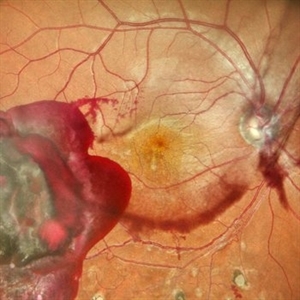
 Large Subhyaloid Hemorrhage
Jul 11 2025 by Jessilla Phou
This is a fundus photograph depicting a large subhyaloid hemorrhage in the mid periphery of the left eye. The patient, a 53-year-old female, presented with a sudden onset of floaters, headache, and blurred vision. The image also demonstrates associated optic disc hemorrhage, vitreous hemorrhage, retinal hemorrhage, and venous tortuosity. Despite the extensive workup performed and the severity of the hemorrhage, no underlying cause was determined.
Photographer: Jessilla Phou
Imaging device: Optos California
Condition/keywords: fundus photograph, optic disc hemorrhage, retinal hemorrhage, venous tortuosity, vitreous hemorrhage
-
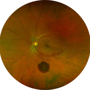
 Prepapillary Vascular Loop
Jul 4 2025 by KANWALJEET HARJOT MADAN, M.S. (Ophthalmology); FAICO (Vitreous - Retina)
This is the fundus picture of right eye of a young 32 years female depicting pre papillary vascular loop. A prepapillary vascular loop is a congenital anomaly of the optic disc that presents as an elevated and twisted bundle of vessels projecting into the vitreous cavity. It is a benign condition, usually unilateral but can be bilateral. It is asymptomatic and discovered during routine eye examination. This anomaly can sometimes cause complications like branch retinal artery occlusion, vitreous hemorrhage, or sub retinal hemorrhage.
Photographer: Dr. Kanwaljeet Harjot Madan, Thind Eye Hospital, Jalandhar City (Punjab) INDIA.
Imaging device: Zeiss Fundus Camera
Condition/keywords: branch retinal artery occlusion (BRAO), optic disc, Prepapillary Vascular Loop, SUB RETINAL HEMORRHAGE, Vitreous hemorrhage
-
 Scleral Rupture
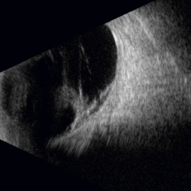
May 9 2025 by Gustavo Uriel Fonseca Aguirre
This B-mode longitudinal ultrasound scan reveals dense vitreous hemorrhage, subretinal fluid, annular choroidal detachment, and scleral wall discontinuity with adjacent scleral folds. These findings indicate severe ocular trauma with probable scleral rupture and multi-compartment involvement.
Photographer: Gustavo U. Fonseca Aguirre, Hospital Conde de Valenciana, Ciudad de México
Condition/keywords: ocular trauma, scleral rupture
-
 Traumatic T
May 5 2025 by Gustavo Uriel Fonseca Aguirre
This B-mode axial ultrasound scan reveals vitreous hemorrhage, a folded retinal detachment, and sub-Tenon’s fluid extending into the optic nerve sheath, forming the characteristic 'T-sign.' These findings are consistent with severe posterior segment trauma secondary to blunt ocular injury.
Photographer: Gustavo U. Fonseca Aguirre, Hospital Conde de Valenciana, Ciudad de México
Condition/keywords: blunt trauma, retinal detachment, T sign
-
 Traumatic Retinal Detachment
May 5 2025 by Gustavo Uriel Fonseca Aguirre
This B-mode longitudinal ultrasound scan over the macular area reveals vitreous hemorrhage, retinal detachment with folding, peripheral annular choroidal detachment, and sub-Tenon's fluid in the setting of blunt ocular trauma. The findings indicate severe posterior segment disruption with multi-compartment involvement.
Photographer: Gustavo U. Fonseca Aguirre, Hospital Conde de Valenciana, Ciudad de México
Condition/keywords: blunt trauma, Retinal Detachment
-
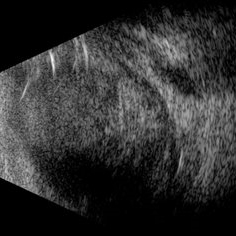
Vitreous Waltz vs Retinal Rigidity
Apr 18 2025 by Gustavo Uriel Fonseca Aguirre
B-mode dynamic ultrasound of an eye with vitreous hemorrhage shows hyaloid traction inducing retinal detachment in diabetic retinopathy. The video clearly delineates all anatomical compartments: vitreous, subhyaloid, and subretinal spaces. Characteristic movement patterns are observed - the vitreous demonstrates smooth, wide excursions while the detached retina shows shorter, stiffer motions -confirming tractional pathology.
Condition/keywords: diabetic retinopathy, retinal detachment
-
 Traumatic Hemorrhage
Apr 17 2025 by Virginia Gebhart
60 year old male with vitreous and sub hyaloid hemorrhage from being hit in the eye. No holes, tears, or detachment. Will observe closely, if no improvement will consider surgical repair. Treated melanoma s/p brachytherapy in 2008.
Photographer: Virginia Gebhart, Retina Consultants of Carolina
Imaging device: Optos California
Condition/keywords: blunt trauma, sub hyaloid hemorrhage, vitreous hemorrhage
-
 Advanced Proliferative Diabetic Retinopathy
Apr 9 2025 by Gustavo Uriel Fonseca Aguirre
B-mode ultrasound of a patient with long-standing poorly controlled diabetes demonstrates characteristic findings of advanced proliferative diabetic retinopathy. The examination reveals moderate vitreous hemorrhage appearing as diffuse hyperechoic opacities throughout the vitreous cavity, along with a posterior hyaloid membrane densely infiltrated by hemorrhagic material, showing irregular thickening and increased reflectivity. A mild subhyaloid hemorrhage is visible as a subtle hyphema-like space anterior to the retinal surface. The study documents a total tractional retinal detachment, evidenced by rigid retinal folds with clear insertion points of vitreous strands, accompanied by a significant subretinal hemorrhage seen as a prominent hyperechoic collection beneath the elevated retina. These findings collectively illustrate the severe vitreoretinal interface pathology characteristic of end-stage diabetic eye disease, with predominant tractional components and distinct echographic stratification of hemorrhagic layers - from anterior vitreous involvement to deeper subretinal blood accumulation.
Photographer: Gustavo U. Fonseca Aguirre, Hospital Conde de Valenciana, Ciudad de México
Condition/keywords: diabetic retinopathy, tractional retinal detachment, Vitreous hemorrhage
-
 Actively Bleeding NVE
Apr 1 2025 by Jordyn Beckman
47 year old woman presented with actively bleeding NVE temporally on exam with complaints of foggy vision and floaters.
Photographer: Jordyn Beckman, Retina Consultants of Carolina, P.A.
Imaging device: Optos California
Condition/keywords: active bleeding, Elevated retinal neovascularization, vitreous hemorrhage
-
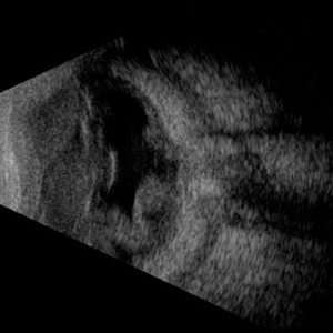
 Macular Hole in Tractional Retinal Detachment
Apr 1 2025 by Gustavo Uriel Fonseca Aguirre
B-scan findings in a diabetic patient reveal vitreous hemorrhage, blood-soaked hyaloids, and tractional retinal detachment with an associated macular hole in the posterior pole.
Photographer: Gustavo U. Fonseca Aguirre, Hospital Conde de Valenciana, Ciudad de México
Condition/keywords: macular hole, tractional retinal detachment
-
 ROP
ROP
Mar 26 2025 by Korey Starkey
9 month old patient presents today with Retinopathy of Prematurity in both eyes. Patient was born at gestational age of 25 weeks 2 days, 940g. Left eye presents with vitreoretinal traction and peripheral VH with regressed stage 3 and persistent stage 2 disease.
Photographer: Korey Starkey
Imaging device: Optos
Condition/keywords: retinopathy of prematurity stage 2, rop, stage 3, vitreoretinal traction, vitreous hemorrhage
-
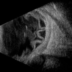
 Hyaloid Butterfly
Mar 13 2025 by Gustavo Uriel Fonseca Aguirre
Axial ultrasound showing a phakic eye with vitreous hemorrhage, hyaloids impregnated with blood, hyalochisis (butterfly-shaped), subhyaloid hemorrhage, and retinal tractions involving the macular area.
Photographer: Gustavo U. Fonseca Aguirre, Hospital Conde de Valenciana, Ciudad de México
Condition/keywords: Hyaloschisis, Sub hyaloid haemorrhage, Vitreous hemorrhage
-
 Retinal Detachment Secondary to Anomalous PVD
Mar 13 2025 by Fabricio Dolores
This color wide-field clinical image depicts the right eye of a female patient who experienced a sudden loss of vision one month earlier. She was initially diagnosed with a vitreous hemorrhage and managed with conservative treatment. Upon presentation to our institute one month later, a superior rhegmatogenous retinal detachment was identified, extending across the 12 o’clock meridian. This was accompanied by an inferior vitreous hemorrhage and a solitary superior retinal lesion located at M11 in the superior triangle of the ora serrata, in alignment with Lincoff's second law.
Photographer: Fabricio Dolores-Villanueva, MD
Imaging device: Nidek Mirante
Condition/keywords: Retinal Detachment
-
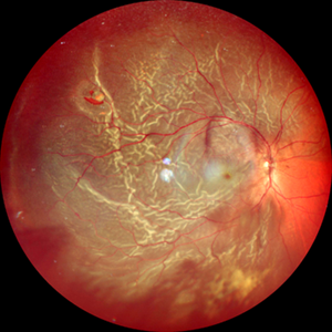
 Papillary Hemorrhage and Associated Vitreous Hemorrhage
Mar 3 2025 by Juan J. Prados-Carmona
Juxtapapillary thrombosis with retinal hemorrhage and associated vitreous hemorrhage localized in the posterior pole.
Photographer: Juan J. Prados-Carmona; Hospital Universitario Reina Sofía (Córdoba), España.
Condition/keywords: hemorrhage
-
 Choroidal Melanoma Masquerading as PEHCR
Mar 3 2025 by Tejaswita Verma
A 65 year old diabetic male presented with large nasal retinal mass giving the appearance of organized dehaemoglobinized subretinal hemorrhage with breakthrough vitreous hemorrhage , with 6/6P vision. Enucleation specimen showed histopathology confirmed choroidal melanoma.
Photographer: DR. TEJASWITA VERMA
Imaging device: MIRANTE
Condition/keywords: vitreous hemorrhage
-
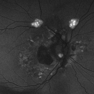 Astrocytic Hamartoma
Feb 27 2025 by Daniel Davis, OCT-C
Fundus autofluorescence photo of 55-year-old female with astrocytic hamartoma in association with tuberous sclerosis. No treatment options available, benign. Other findings include; Posterior Vitreous Detachment, Vitreous Hemorrhage, Hereditary Retinal Dystrophy, Vitreous Opacities, Hypertensive Retinopathy.
Photographer: Daniel Davis, OCT-C
Imaging device: Optos California
Condition/keywords: astrocytic hamartoma, fundus autofluorescence (FAF)
-
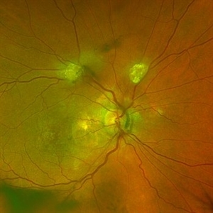 Astrocytic Hamartoma
Feb 27 2025 by Daniel Davis, OCT-C
Color fundus photo of 55-year-old female with Astrocytic Hamartoma in association with tuberous sclerosis. No treatment options available, benign. Other findings include; Posterior Vitreous Detachment, Vitreous Hemorrhage, Hereditary Retinal Dystrophy, Vitreous Opacities, Hypertensive Retinopathy.
Photographer: Daniel Davis, OCT-C
Imaging device: Optos California
Condition/keywords: color fundus photograph
-
 Firework Injury
Feb 13 2025 by Virginia Gebhart
44 year old male presented New Year's Day for trauma after fireworks injury. Choroidal rupture temporal macula, inferior vitreous hemorrhage, and extensive RPE changes in the macula. Significant improvement since initial presentation. Limited central vision, guarded prognosis due to extensive blunt trauma.
Photographer: Virginia Gebhart, Retina Consultants of Carolina
Imaging device: Optos California
Condition/keywords: blunt trauma, choroidal rupture, commotio retinae, firework injury, secondary glaucoma, subretinal hemorrhage, VH, vitreous hemorrhage
-
 Ciliary Body Melanoma
Feb 12 2025 by Virginia Gebhart
91 year old female with large collar button tumor emanating from the ciliary body with resolving vitreous hemorrhage. Melanoma cells in the AV as well as studded on the entire retina surface. Pt scheduled for enucleation. CT scans of chest and abdomen showed no evidence of metastatic disease.
Photographer: Virginia Gebhart, Retina Consultants of Carolina
Imaging device: Optos California
Condition/keywords: asteroid hyalosis, ciliary body mass, ciliary body melanoma, vitreous hemorrhage
-
 Diabetic Vitreous Hemorrhage
Feb 3 2025 by Hollie Sanders
61 year-old male with a history of type two DM. Per MD, Sub-Retinal, Pre-Retinal, and Vitreous Hemorrhages. Denies any treatment in the past. Treatment initiated in clinic.
Photographer: Hollie Sanders, Tennessee Retina, Nashville, Tennessee
Imaging device: OPTOS Silverstone Fundus camera
Condition/keywords: diabetic vitreous hemorrhage
-
 Dislocated Lens
Jan 30 2025 by Kimberly Wakester
Fundus photograph of a 37-year-old man with an anteriorly dislocated lens in the left eye. The natural lens has displaced anteriorly in the AC secondary to trauma to the eye. There is also a Macular hole present with vitreous hemorrhage. Patient was recommended to proceed with lensectomy, iris repair and MH repair in the left eye.
Photographer: Kimberly Wakester, COA
Imaging device: Topcon TRC-50DX
Condition/keywords: dislocated lens, iridodialysis
-
 Macular Hole
Jan 30 2025 by Kimberly Wakester
Fundus photograph of a 37-year-old man with an anteriorly dislocated lens in the left eye. The natural lens has displaced anteriorly in the AC secondary to trauma to the eye. There is also a Macular hole present with vitreous hemorrhage. Patient was recommended to proceed with lensectomy, iris repair and MH repair in the left eye.
Photographer: Kimberly Wakester, COA
Imaging device: Optos California
Condition/keywords: dislocated lens, macular hole, vitreous hemorrhage
-
 Choroidal Melanoma Masquerading as Subretinal Hemorrhage With Breakthrough VH
Jan 23 2025 by Tejaswita Verma
A 65 year old diabetic male presented with large nasal retinal mass giving the appearance of organized dehaemoglobinized subretinal hemorrhage with breakthrough vitreous hemorrhage , with 6/6P vision. Enucleation specimen showed histopathology confirmed choroidal melanoma.
Photographer: DR. TEJASWITA VERMA
Imaging device: MIRANTE
 Loading…