Search results (59 results)
-
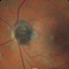
 ERMageddon - Wrinkle in the Space-time Fabric of Macula
Oct 29 2025 by SHRADDHA RAJ SHRIVASTAVA
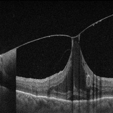
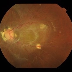
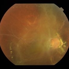
38 year old female with Epiretinal Membrane (ERM) over macula, post laser barrage for multiple symptomatic Horse-shoe Tears (HSTs) and Lattice Degenerations (seen on wide-field image). Posterior pole revealed tilted disc with peripapillary atrophy. There is thick opaque epiretinal membrane obscuring the underlying superior arcade vessels and causing foveal ectopia with distortion of perimacular vasculature. Patient was planned for Right Eye pars plana vitrectomy for ERM peeling.
Photographer: Dr. Shraddha Raj Shrivastava
Imaging device: Nidek Mirante SLO/OCT (Confocal scanning/Spectral domain OCT
Condition/keywords: BARRAGE LASER, ectopic fovea, epiretinal membrane (ERM), horseshoe tear, lattice degeneration, vitreomacular traction (VMT)
-
 ERMageddon - Wrinkle in the Space-time Fabric of Macula
Oct 29 2025 by SHRADDHA RAJ SHRIVASTAVA
38 year old female with Epiretinal Membrane (ERM) over macula, post laser barrage for multiple symptomatic Horse-shoe Tears (HSTs) and Lattice Degenerations. Posterior pole revealed tilted disc with peripapillary atrophy. There is thick opaque epiretinal membrane obscuring the underlying superior arcade vessels and causing foveal ectopia with distortion of perimacular vasculature. Patient was planned for Right Eye pars plana vitrectomy for ERM peeling.
Photographer: Dr. Shraddha Raj Shrivastava
Imaging device: Nidek Mirante SLO/OCT (Confocal scanning/Spectral domain OCT
Condition/keywords: ectopic fovea, epiretinal membrane (ERM), ERM, horseshoe tear, vitreomacular traction (VMT)
-
 Vitreomacular Adhesion Showcasing a Microaneurysm and a Subhyaloid Hemorrhage
Jan 3 2025 by Drew Mitchell
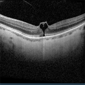
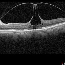
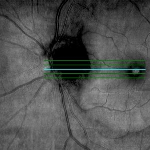
A vertical OCT 1 line raster scan positioned slightly inferomacula to document the subhyaloid hemorrhage. Hyper reflective Oval indicating Microaneurysm.
Photographer: Drew Mitchell, OCT-C
Imaging device: Zeiss Cirrus 5000
Condition/keywords: diabetic macular edema, microaneurysms, retinal microaneurysms, subhyaloid hemorrhage, subretinal fluid, vitreomacular adhesion, vitreomacular traction (VMT)
-
 Eye Heart You
Jul 23 2024 by Ashley Phillips
VMT causing macular hole.
Photographer: Ashley Phillips
Imaging device: Zeiss 5000
Condition/keywords: vitreomacular traction (VMT)
-
 Ozurdex Implant on OCT
Jul 18 2024 by Reghan Knight-Rabley
OCT of 66 year old woman with history of BRVO with macular edema, showing migration of Ozurdex implant.
Photographer: Reghan Knight-Rabley, OMA, Retina Specialists of Michigan
Condition/keywords: branch retinal vein occlusion (BRVO), BRVO, intravitreal, ozurdex, Ozurdex implant, vitreomacular traction (VMT)
-
 Vitreomacular traction in a case of advanced exudative AMD
Oct 16 2022 by T. P . VIGNESH, MBBS,MS
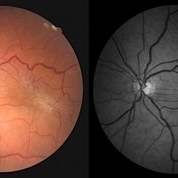
SD-OCT of 70 year old man revealing scarred CNVM and VMT .
Photographer: Shivanath
Imaging device: Heidelberg Spectralis
Condition/keywords: vitreomacular traction (VMT)
-
 Proliferative Diabetic Retinopathy
Aug 16 2022 by Donnie Willis
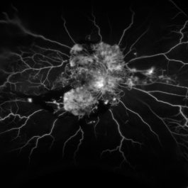
51 yo female. Uncontrolled Diabetes. Active PDR.
Photographer: Donnie Willis, Tennessee Retina
Imaging device: Optos
Condition/keywords: capillary dropouts, Diabetes, FA, Optos, proliferative diabetic retinopathy (PDR), vitreomacular traction (VMT)
-
 Vitreomacular traction
Jun 23 2022 by T. P . VIGNESH, MBBS,MS
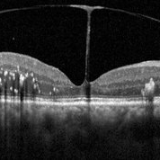
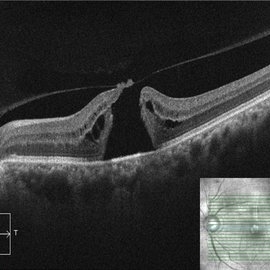
SD-OCT of RE reveals Vitreomacular traction resembling bow and arrow and diabetic macular edema with intraretinal hard exudates in a 60 year old female patient with Moderate NPDR .
Imaging device: Heidelberg Spectralis
Condition/keywords: vitreomacular traction (VMT)
-
 Vitreomacular Traction
Jun 15 2022 by Zach Seim
Optical Coherence Tomography (OCT) of a 69 year old male with Vitreomacular Traction affecting his right eye. Patient was referred to this office for Choroidal Melanoma in his right eye in May 2021. The patient was treated with Brachytherapy in July 2021 and this OCT was taken at a follow-up appointment in May 2022. Patient's vision was 20/30-2 at the time this OCT was taken. Patient states that his vision was better since his last visit, and that he sees floaters occasionally.
Photographer: Zach Seim
Imaging device: Heidelberg Spectralis
Condition/keywords: heidelberg spectralis, OD, optical coherence tomography (OCT), right eye, subretinal fluid, vitreomacular adhesion, vitreomacular interface disorders, vitreomacular traction (VMT)
-
 Eiffel tower within the eye
Mar 31 2022 by Bhavani Sankaran, MS (Ophthalmology)
OCT of left eye of a 68 year old female patient who presented with complaints of metamorphopsia. Image depicts vitreomacular traction. BCVA OS 20/40.
Photographer: Dr. Bhavani Sankaran, MS, Aravind Eye Hospital, Madurai
Imaging device: Heidelberg Spectralis
Condition/keywords: vitreomacular interface disorders, vitreomacular traction (VMT)
-
 OCT Evidence of VMT Resulting in Full Thickness Macular Hole
Dec 24 2020 by Deepak Bhojwani, MS
OCT image of a patient (with past history of focal VMT ) progressing to full thickness macular hole. Note the posterior hyaloid attachment over the torn edges of fovea.
Photographer: DEEPAK BHOJWANI
Condition/keywords: full thickness macular hole, optical coherence tomography (OCT), vitreomacular traction (VMT)
-
 Vitreo-Macular Traction Syndrome
Sep 1 2020 by J. Sebag, MD, FACS, FRCOphth, FARVO
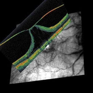
Combined OCT and Scanning laser ophthalmoscopy demonstrate separation of full-thickness posterior vitreous cortex (no vitreoschisis), but persistent adhesion centrally with significant detachment of the fovea. [from Sebag J: Vitreous – in Health & Disease (J. Sebag, ed.) Springer, New York, 2014; image © Springer Nature, reprinted with permission]
Condition/keywords: vitreomacular traction (VMT)
-
 Enucleated Eye with Retinal Atrophy, Vitreomacular Traction and Keratoconus
May 18 2020 by McGill University Health Centre
This enucleation specimen shows areas of retinal atrophy (*) and areas of vitreomacular traction (arrow). This specimen also demonstrates keratoconus: a degenerative disorder of the eye in which the cornea thins and distorts into a pronounced conical shape (arrowhead). The keratoconus and vitreomacular tractions are unrelated.
Condition/keywords: atrophy, keratoconus, vitreomacular traction (VMT)
-
 Combined Hamartoma of Retina and RPE (OS)
Jan 18 2020 by Haider Ali
Fundus photograph of 14-year-old girl with hand movements vision in both eyes. No history of any systemic disease.
Photographer: Haider Ali Chaudhry, Madinah Teaching Hospital, Faisalabad
Condition/keywords: combined hamartoma, epiretinal membrane (ERM), epiretinal membrane formation, neurofibromatosis, vitreomacular traction (VMT)
-
 Combined Hamartoma of Retina and RPE (OD)
Jan 18 2020 by Haider Ali
Fundus photograph of 14-year-old girl with hand movements vision in both eyes. No history of any systemic disease.
Photographer: Haider Ali Chaudhry, Madinah Teaching Hospital, Faisalabad
Condition/keywords: combined hamartoma, epiretinal membrane (ERM), epiretinal membrane formation, vitreomacular traction (VMT)
-
 Spontaneous Resolution of Vitreomacular Traction
Dec 28 2019 by Anfisa Ayalon, MD
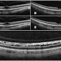
A 42-year-old highly myopic patient presented to the retina clinic with complains of metamorphopsia in his RE for the past months. SD-OCT revealed RE vitreomacular traction with stage 1 macular hole (A-B) and flat myopic retinoschisis (C). Visual acuity in the same eye was 6/30. After 3 weeks of follow up spontaneous resolution of vitreomacular traction was seen with full resolution of the symptoms. (D-E). Visual acuity remained without significant change.
Photographer: Anfisa Ayalon,MD., Meir Medical Center, Kfar Saba, Israel.
Condition/keywords: flat retinoschisis, macular hole, myopia, stage 1 macular hole, vitreomacular traction (VMT)
-
 Spontaneous Resolution of Vitreomacular Traction
Dec 28 2019 by Anfisa Ayalon, MD
A 42-year-old highly myopic patient presented to the retina clinic with complains of metamorphopsia for the past months. SD-OCT revealed RE vitreomacular traction with stage 1 macular hole (pictures from the left). After 3 weeks of follow up spontaneous resolution of vitreomacular traction (pictures from the right) was seen with full resolution of the symptoms.
Photographer: Anfisa Ayalon, MD., Meir Medical Center, Kfar Saba, Israel.
Condition/keywords: macular hole, myopia, spontaneous resolution, stage 1 macular hole, vitreomacular traction (VMT)
-
 Epiretinal Membrane
Sep 26 2018 by Andrea Arriola-Lopez, MD MSc
45-year-old woman, BCVA 20/80 OS. Indirect ophthalmoscopy revealed epiretinal membrane, macular folds and traction. PPV was scheduled.
Photographer: Lourdes Guambo MD, Centro Oftalmológico León, UFM.
Condition/keywords: epiretinal membrane (ERM), vitreomacular interface disorders, vitreomacular traction (VMT)
-
 Melanocytoma and Vitreomacular Traction Syndrome OCT Fundus Image
Mar 19 2018 by Nelson Antonio Segovia Rodríguez, MD
OCT fundus image of an 68-year-old male with a optic dis melanocytoma and associated vitreomacular traction syndrome.
Photographer: Nelson Segovia, private practice
Imaging device: Zeiss Cirrus
Condition/keywords: melanocytoma, vitreomacular traction (VMT)
-
 Melanocytoma and Vitreomacular Traction Syndrome Fundus Color Image
Mar 19 2018 by Nelson Antonio Segovia Rodríguez, MD
Color fundus image of an 68-year-old male with a optic dis melanocytoma and associated vitreomacular traction syndrome.
Photographer: Nelson Segovia, private practice
Imaging device: Zeiss
Condition/keywords: melanocytoma, optical coherence tomography (OCT), vitreomacular traction (VMT)
-
 Age-Related-Maculopathy with Advanced Vitreo Macular Traction in Right Eye
Feb 7 2018 by Ogugua Ndubuisi Okonkwo, MD, FRCS (Edin), FASRS
Right eye Fundus photograph of a 73-year-old woman with age related maculopathy, progressive drusenoid deposits and progressively worsening Vitreomacular Traction (VMT) monitored over a 10 year duration. Requiring surgical release of VMT.
Photographer: Okonkwo Ogugua, Eye Foundation Retina Institute . Lagos
Condition/keywords: drusenoid deposit, vitreomacular adhesion, vitreomacular traction (VMT)
-
 Age-Related-Maculopathy and Vitreo Macular Adhesion in Left Eye
Feb 7 2018 by Ogugua Ndubuisi Okonkwo, MD, FRCS (Edin), FASRS
Left eye Fundus photograph of a 73-year-old woman with age related maculopathy , progressive drusenoid deposits and Vitreomacular Adhesion (VMA) monitored over a 10 year duration.
Photographer: Okonkwo Ogugua, Eye Foundation Retina Institute . Lagos
Condition/keywords: drusenoid deposit, vitreomacular traction (VMT)
-
 Pre-Op-Fern-Like-Appearance-of-VMT
Feb 7 2018 by Ogugua Ndubuisi Okonkwo, MD, FRCS (Edin), FASRS
Right eye pre operative OCT of a 73-year-old woman with age related maculopathy and progressively worsening Vitreomacular Traction (VMT) , requiring surgical release of VMT. Fern like VMT is seen and undulating RPE layer showing the drusenoid deposits.
Photographer: Oreoluwa , Eye Foundation Retina Institute . Lagos
Condition/keywords: drusenoid deposit, vitreomacular traction (VMT)
-
 Right Eye One month Post Operative VMT Relaease
Feb 7 2018 by Ogugua Ndubuisi Okonkwo, MD, FRCS (Edin), FASRS
Right eye one month post-operative OCT showing VMT release with return of the foveal depression and good vision. There is marked reduction in intra retina schisis and preserved outer retina layers.
Photographer: Oreoluwa, Eye Foundation Retina Institute . Lagos
Condition/keywords: drusenoid deposit, vitreomacular traction (VMT)
-
 PED and VMA-LE
Feb 7 2018 by Ogugua Ndubuisi Okonkwo, MD, FRCS (Edin), FASRS
Left eye OCT of same 73-year-old female showing drusenoid PED and Vitreomacular Adhesion .
Photographer: Oreoluwa, Eye Foundation Retina Institute . Lagos
Condition/keywords: drusenoid deposit, vitreomacular traction (VMT)
 Loading…