Search results (249 results)
-
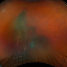 Retinal Tear w/VH
Aug 22 2025 by Virginia Gebhart
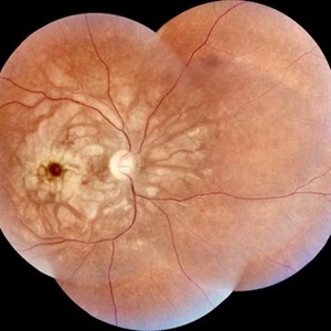
69 year old male referred for sudden vision loss. Difficult view secondary to VH. Ultrasound and photos show small break with clotting, heavy amount of layered blood inferior and scattered IRH's. Cryotherapy performed to seal current tear, will give VH time to clear on its own. Pt takes Eliquis twice a day.
Photographer: Virginia Gebhart, Retina Consultants of Carolina
Imaging device: Optos California
Condition/keywords: cryo-retinal tear, horseshoe tear, tear, vitreous hemorrhage
-
 Sub-ILM Hemorrhage
Aug 6 2025 by Virginia Gebhart
28 year old female with sub-ILM hemorrhage and questionable cotton wool spot. Pt states sudden vision loss after being hit in the eye with laser lights at a nightclub. Laser lights also damaged the camera in her iPhone. Will give hemorrhage more time to clear on its own, will discuss treatment options if no improvement at next visit.
Photographer: Virginia Gebhart, Retina Consultants of Carolina
Imaging device: Topcon TRC 50DX
Condition/keywords: retinal hemorrhage, sub ILM hemorrhage
-
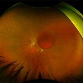
 Traction in Progression
Aug 6 2025 by Claudio Brancato, MD
This image captures stage 4A Retinopathy of Prematurity, showing partial retinal detachment sparing the macula. The elevated retina and fibrous ridge indicate tractional forces secondary to extraretinal neovascularization. A striking representation of disease evolution, poised between reversibility and vision loss.
Photographer: Gregorio Lo Giudice, ARNAS Civico Hospital, Palermo, Italy
Imaging device: RETCAM 3 (enhanced via IA)
Condition/keywords: retinopathy of prematurity
-
 Hemorrhagic Retinal Arterial Macroaneurysm
Jul 4 2025 by Julian Navarro Saucedo
Fundus photograph of a 52-year-old woman with sudden vision loss showing a hemorrhagic retinal arterial macroaneurysm.
Photographer: Julian Navarro, Tecnologico de Monterrey, Escuela de Medician y Ciencias de la Salud.
Imaging device: VISUCAM 524 ZEISS
Condition/keywords: macroaneurysm
-
 Purtscher-like Retinopathy in Preeclampsia
Jun 28 2025 by Sriharanathan Poopalaratnam, MD,FRCS
A 24-year-old female, 6 weeks post-emergency cesarean section (LSCS), with a history of pregnancy-induced hypertension (PIH), presented with acute, profound, bilateral painless vision loss of 2 days’ duration
Photographer: Yattiwarra
Condition/keywords: preeclampsia, Purtscher like
-
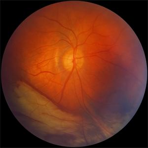
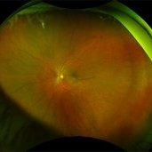
 Aggressive Posterior Retinopathy of Prematurity (APROP)
May 16 2025 by KANWALJEET HARJOT MADAN, M.S. (Ophthalmology); FAICO (Vitreous - Retina)
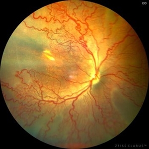
This is the fundus picture of right eye of a premature neonate depicting Aggressive Posterior Retinopathy of Prematurity (APROP). It is a severe rapidly progressing form of retinopathy that can lead to vision loss and blindness. It requires prompt diagnosis and treatment in the form of anti-VEGF agents and laser photocoagulation.
Photographer: Dr. Kanwaljeet Harjot Madan, Thind Eye Hospital, Jalandhar City (Punjab) INDIA.
Imaging device: Zeiss Clarus
Condition/keywords: Oxygen Exposure, retinopathy of prematurity (ROP)
-
 LCA Type 1
Apr 10 2025 by Joshua Friedman
LCA Type 1 (GUCY2D) demonstrating the hallmark discordance between severe vision loss (light perception) in a 10-year-old female and a near-normal fundus appearance in childhood, typical of guanylate cyclase dysfunction.
Photographer: Stephen Tsang, MD, PhD
Condition/keywords: Leber Congenital Amaurosis (LCA)
-
 Cilioretinal Artery Occlusion
Dec 12 2024 by César Adrián Gómez Valdivia, MD
Cilioretinal artery occlusion found in a 25 year-old male patient with history of monocular, sudden, painless vision loss.
Photographer: @eyemissu2
Imaging device: TOPCON TRC-50DX
Condition/keywords: cilioretinal artery occlusion, oclussion
-
 Atypical RP with Typhoid Retinitis Sequelae with Old CRAO
Dec 5 2024 by Tejaswita Verma
FAF of a 20 year old female who presented with 2 months history of sudden painless vision loss, bilaterally light perception vision, s/o presumed atypical RP, bilateral old CRAO with typhoid retinitis sequelae.
Photographer: DR. TEJASWITA VERMA
Imaging device: MIRANTE
Condition/keywords: CRAO, retinitis pigmentosa, typhoid fever
-
 MIDD (Maternally Inherited Diabetes and Deafness) - Left AF
Nov 30 2024 by John S. King, MD
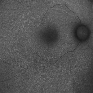
Both right and left eyes have symmetrical ring of mottled hypo/hyper AF around the fovea and disc. The HyperAF areas correspond to RPE deposits on OCT as well as areas of blockage on FA, and drusenoid deposits seen on fundus photos 57 yo WF referred for AMD vs Pattern Dystrophy that was diagnosed 10 years ago. Reported some slow progressive vision loss in both eyes for distance and near. Denies nyctalopia or hemeralopia. Background medical history includes HTN, CVD, and DM. No family history of eye problems. Denied pentosan use. Anterior segment showed moderate cataracts (OD>OS). Posterior segment exam showed macular changes and mild NPDR. The macular appearance showed a symmetrical, paramacular ring of fleck-like drusenoid material with some faint focal areas of RPE hyperplasia. Fundus Photos, AF, OCT were performed as well as a gene test. Further questioning showed revealed that her mother and maternal grandmother had both diabetes mellitus and sensorineural hearing loss. The patient developed diabetes in her teens, and some high frequency hearing loss in her early twenties. She had not had a previous genetic test or diagnosis of MIDD. Gene testing is pending for the mitochondrial component. Invitae's retinal panel, which does not include mitochondrial disorders, only showed a variant of uncertain significance, HMCN1. I discussed this case with Dr. Freund, and it is similar to a the case report : Inoue M, Kiss S, Freund KB. MACULAR PIGMENT RINGS AS THE PRESENTING FINDING OF MITOCHONDRIAL MYOPATHY, ENCEPHALOPATHY, LACTIC ACIDOSIS, AND STROKELIKE EPISODES. Retin Cases Brief Rep. 2015 Fall;9(4):260-4. doi: 10.1097/ICB.0000000000000182. PMID: 26200388.
Photographer: Grace Melton and Carley Gunn
Imaging device: Clarus
Condition/keywords: Macular Dystrophy, Maternally Inherited Diabetes and Deafness, MIDD, Mitochondrial Disorder
-
 MIDD (Maternally Inherited Diabetes and Deafness) - Right AF
Nov 30 2024 by John S. King, MD
Both right and left eyes have symmetrical ring of mottled hypo/hyper AF around the fovea and disc. The HyperAF areas correspond to RPE deposits on OCT as well as areas of blockage on FA, and drusenoid deposits seen on fundus photos. Disc drusen in right eye present as HyperAF spot 57 yo WF referred for AMD vs Pattern Dystrophy that was diagnosed 10 years ago. Reported some slow progressive vision loss in both eyes for distance and near. Denies nyctalopia or hemeralopia. Background medical history includes HTN, CVD, and DM. No family history of eye problems. Denied pentosan use. Anterior segment showed moderate cataracts (OD>OS). Posterior segment exam showed macular changes and mild NPDR. The macular appearance showed a symmetrical, paramacular ring of fleck-like drusenoid material with some faint focal areas of RPE hyperplasia. Fundus Photos, AF, OCT were performed as well as a gene test. Further questioning showed revealed that her mother and maternal grandmother had both diabetes mellitus and sensorineural hearing loss. The patient developed diabetes in her teens, and some high frequency hearing loss in her early twenties. She had not had a previous genetic test or diagnosis of MIDD. Gene testing is pending for the mitochondrial component. Invitae's retinal panel, which does not include mitochondrial disorders, only showed a variant of uncertain significance, HMCN1. I discussed this case with Dr. Freund, and it is similar to a the case report : Inoue M, Kiss S, Freund KB. MACULAR PIGMENT RINGS AS THE PRESENTING FINDING OF MITOCHONDRIAL MYOPATHY, ENCEPHALOPATHY, LACTIC ACIDOSIS, AND STROKELIKE EPISODES. Retin Cases Brief Rep. 2015 Fall;9(4):260-4. doi: 10.1097/ICB.0000000000000182. PMID: 26200388.
Photographer: Grace Melton and Carley Gunn
Imaging device: Clarus
Condition/keywords: Macular Dystrophy, Maternally Inherited Diabetes and Deafness, MIDD, Mitochondrial Disorder
-
 MIDD (Maternally Inherited Diabetes and Deafness) - Left FP
Nov 30 2024 by John S. King, MD
Both the right and left Eye have fairly symmetrical, extrafoveal drusenoid-like flecks and focal and faint areas of RPE hyperplasia (in addition to mild NPDR and PPA) 57 yo WF referred for AMD vs Pattern Dystrophy that was diagnosed 10 years ago. Reported some slow progressive vision loss in both eyes for distance and near. Denies nyctalopia or hemeralopia. Background medical history includes HTN, CVD, and DM. No family history of eye problems. Denied pentosan use. Anterior segment showed moderate cataracts (OD>OS). Posterior segment exam showed macular changes and mild NPDR. The macular appearance showed a symmetrical, paramacular ring of fleck-like drusenoid material with some faint focal areas of RPE hyperplasia. Fundus Photos, AF, OCT were performed as well as a gene test. Further questioning showed revealed that her mother and maternal grandmother had both diabetes mellitus and sensorineural hearing loss. The patient developed diabetes in her teens, and some high frequency hearing loss in her early twenties. She had not had a previous genetic test or diagnosis of MIDD. Gene testing is pending for the mitochondrial component. Invitae's retinal panel, which does not include mitochondrial disorders, only showed a variant of uncertain significance, HMCN1. I discussed this case with Dr. Freund, and it is similar to a the case report : Inoue M, Kiss S, Freund KB. MACULAR PIGMENT RINGS AS THE PRESENTING FINDING OF MITOCHONDRIAL MYOPATHY, ENCEPHALOPATHY, LACTIC ACIDOSIS, AND STROKELIKE EPISODES. Retin Cases Brief Rep. 2015 Fall;9(4):260-4. doi: 10.1097/ICB.0000000000000182. PMID: 26200388.
Photographer: Grace Melton and Carley Gunn
Imaging device: Clarus
Condition/keywords: Macular Dystrophy, Maternally Inherited Diabetes and Deafness, MIDD, Mitochondrial Disorder
-
 MIDD (Maternally Inherited Diabetes and Deafness) - Right FP
Nov 30 2024 by John S. King, MD
Both the right and left Eye have fairly symmetrical, extrafoveal drusenoid-like flecks and focal and faint areas of RPE hyperplasia (in addition to mild NPDR and PPA) 57 yo WF referred for AMD vs Pattern Dystrophy that was diagnosed 10 years ago. Reported some slow progressive vision loss in both eyes for distance and near. Denies nyctalopia or hemeralopia. Background medical history includes HTN, CVD, and DM. No family history of eye problems. Denied pentosan use. Anterior segment showed moderate cataracts (OD>OS). Posterior segment exam showed macular changes and mild NPDR. The macular appearance showed a symmetrical, paramacular ring of fleck-like drusenoid material with some faint focal areas of RPE hyperplasia. Fundus Photos, AF, OCT were performed as well as a gene test. Further questioning showed revealed that her mother and maternal grandmother had both diabetes mellitus and sensorineural hearing loss. The patient developed diabetes in her teens, and some high frequency hearing loss in her early twenties. She had not had a previous genetic test or diagnosis of MIDD. Gene testing is pending for the mitochondrial component. Invitae's retinal panel, which does not include mitochondrial disorders, only showed a variant of uncertain significance, HMCN1. I discussed this case with Dr. Freund, and it is similar to a the case report : Inoue M, Kiss S, Freund KB. MACULAR PIGMENT RINGS AS THE PRESENTING FINDING OF MITOCHONDRIAL MYOPATHY, ENCEPHALOPATHY, LACTIC ACIDOSIS, AND STROKELIKE EPISODES. Retin Cases Brief Rep. 2015 Fall;9(4):260-4. doi: 10.1097/ICB.0000000000000182. PMID: 26200388.
Photographer: Grace Melton and Carley Gunn
Imaging device: Clarus
Condition/keywords: Macular Dystrophy, Maternally Inherited Diabetes and Deafness, MIDD, Mitochondrial Disorder
-
 MIDD (Maternally Inherited Diabetes and Deafness) - OCT OD
Nov 30 2024 by John S. King, MD
OCT shows mild RPE deposit inferiorly (corresponds to area of FA blockage and HyperAF) and a focal area of iRORA with loss of EZ more superiorly (possibly due to regression of RPE deposit). No choroidal thickening (like in pachychoroid pigment epitheliopathy or cscr) 57 yo WF referred for AMD vs Pattern Dystrophy that was diagnosed 10 years ago. Reported some slow progressive vision loss in both eyes for distance and near. Denies nyctalopia or hemeralopia. Background medical history includes HTN, CVD, and DM. No family history of eye problems. Denied pentosan use. Anterior segment showed moderate cataracts (OD>OS). Posterior segment exam showed macular changes and mild NPDR. The macular appearance showed a symmetrical, paramacular ring of fleck-like drusenoid material with some faint focal areas of RPE hyperplasia. Fundus Photos, AF, OCT were performed as well as a gene test. Further questioning showed revealed that her mother and maternal grandmother had both diabetes mellitus and sensorineural hearing loss. The patient developed diabetes in her teens, and some high frequency hearing loss in her early twenties. She had not had a previous genetic test or diagnosis of MIDD. Gene testing is pending for the mitochondrial component. Invitae's retinal panel, which does not include mitochondrial disorders, only showed a variant of uncertain significance, HMCN1. I discussed this case with Dr. Freund, and it is similar to a the case report : Inoue M, Kiss S, Freund KB. MACULAR PIGMENT RINGS AS THE PRESENTING FINDING OF MITOCHONDRIAL MYOPATHY, ENCEPHALOPATHY, LACTIC ACIDOSIS, AND STROKELIKE EPISODES. Retin Cases Brief Rep. 2015 Fall;9(4):260-4. doi: 10.1097/ICB.0000000000000182. PMID: 26200388.
Photographer: Grace Melton and Carley Gunn
Imaging device: Zeiss Cirrus
Condition/keywords: Macular Dystrophy, Maternally Inherited Diabetes and Deafness, MIDD, Mitochondrial Disorder
-
 MIDD (Maternally Inherited Diabetes and Deafness) - OCT OS
Nov 30 2024 by John S. King, MD
Magnified section of radial scan through the left eye showing a focal nodular RPE deposit that corresponds to a focal drusenoid deposit in temporal macula, that HypoFLs and HyperAFs. Choroid not significantly thickened or thinned, and the nodular thickening may be just above a large outer choroid vessel?) 57 yo WF referred for AMD vs Pattern Dystrophy that was diagnosed 10 years ago. Reported some slow progressive vision loss in both eyes for distance and near. Denies nyctalopia or hemeralopia. Background medical history includes HTN, CVD, and DM. No family history of eye problems. Denied pentosan use. Anterior segment showed moderate cataracts (OD>OS). Posterior segment exam showed macular changes and mild NPDR. The macular appearance showed a symmetrical, paramacular ring of fleck-like drusenoid material with some faint focal areas of RPE hyperplasia. Fundus Photos, AF, OCT were performed as well as a gene test. Further questioning showed revealed that her mother and maternal grandmother had both diabetes mellitus and sensorineural hearing loss. The patient developed diabetes in her teens, and some high frequency hearing loss in her early twenties. She had not had a previous genetic test or diagnosis of MIDD. Gene testing is pending for the mitochondrial component. Invitae's retinal panel, which does not include mitochondrial disorders, only showed a variant of uncertain significance, HMCN1. I discussed this case with Dr. Freund, and it is similar to a the case report : Inoue M, Kiss S, Freund KB. MACULAR PIGMENT RINGS AS THE PRESENTING FINDING OF MITOCHONDRIAL MYOPATHY, ENCEPHALOPATHY, LACTIC ACIDOSIS, AND STROKELIKE EPISODES. Retin Cases Brief Rep. 2015 Fall;9(4):260-4. doi: 10.1097/ICB.0000000000000182. PMID: 26200388.
Photographer: Grace Melton and Carley Gunn
Imaging device: Zeiss Cirrus
Condition/keywords: Macular Dystrophy, Maternally Inherited Diabetes and Deafness, MIDD, Mitochondrial Disorder
-
 MIDD (Maternally Inherited Diabetes and Deafness) - Right FA (4 min)
Nov 30 2024 by John S. King, MD
Both eyes had similar FA findings. There was no dark choroid or signs of leakage. Granular staining around the fovea and disc were present, and the HypoAF areas corresponded to the drusenoid deposits that showed HyperAF. Mild MAs present due to NPDR 57 yo WF referred for AMD vs Pattern Dystrophy that was diagnosed 10 years ago. Reported some slow progressive vision loss in both eyes for distance and near. Denies nyctalopia or hemeralopia. Background medical history includes HTN, CVD, and DM. No family history of eye problems. Denied pentosan use. Anterior segment showed moderate cataracts (OD>OS). Posterior segment exam showed macular changes and mild NPDR. The macular appearance showed a symmetrical, paramacular ring of fleck-like drusenoid material with some faint focal areas of RPE hyperplasia. Fundus Photos, AF, OCT were performed as well as a gene test. Further questioning showed revealed that her mother and maternal grandmother had boith diabetes mellitus and sensorineural hearing loss. The patient developed diabetes in her teens, and some high frequency hearing loss in her early twenties. She had not had a previous genetic test or diagnosis of MIDD. Gene testing is pending for the mitochondrial component. Invitae's retinal panel, which does not include mitochondrial disorders, only showed a variant of uncertain significance, HMCN1. I discussed this case with Dr. Freund, and it is similar to a the case report : Inoue M, Kiss S, Freund KB. MACULAR PIGMENT RINGS AS THE PRESENTING FINDING OF MITOCHONDRIAL MYOPATHY, ENCEPHALOPATHY, LACTIC ACIDOSIS, AND STROKELIKE EPISODES. Retin Cases Brief Rep. 2015 Fall;9(4):260-4. doi: 10.1097/ICB.0000000000000182. PMID: 26200388.
Photographer: Grace Melton and Carley Gunn
Imaging device: Clarus
Condition/keywords: Macular Dystrophy, Maternally Inherited Diabetes and Deafness, MIDD, Mitochondrial Disorder
-
 MIDD (Maternally Inherited Diabetes and Deafness) - Left FA (7 min)
Nov 30 2024 by John S. King, MD
Both eyes had similar FA findings. There was no dark choroid or signs of leakage. Granular staining around the fovea and disc were present, and the HypoAF areas corresponded to the drusenoid deposits that showed HyperAF. Mild MAs present due to NPDR 57 yo WF referred for AMD vs Pattern Dystrophy that was diagnosed 10 years ago. Reported some slow progressive vision loss in both eyes for distance and near. Denies nyctalopia or hemeralopia. Background medical history includes HTN, CVD, and DM. No family history of eye problems. Denied pentosan use. Anterior segment showed moderate cataracts (OD>OS). Posterior segment exam showed macular changes and mild NPDR. The macular appearance showed a symmetrical, paramacular ring of fleck-like drusenoid material with some faint focal areas of RPE hyperplasia. Fundus Photos, AF, OCT were performed as well as a gene test. Further questioning showed revealed that her mother and maternal grandmother had boith diabetes mellitus and sensorineural hearing loss. The patient developed diabetes in her teens, and some high frequency hearing loss in her early twenties. She had not had a previous genetic test or diagnosis of MIDD. Gene testing is pending for the mitochondrial component. Invitae's retinal panel, which does not include mitochondrial disorders, only showed a variant of uncertain significance, HMCN1. I discussed this case with Dr. Freund, and it is similar to a the case report : Inoue M, Kiss S, Freund KB. MACULAR PIGMENT RINGS AS THE PRESENTING FINDING OF MITOCHONDRIAL MYOPATHY, ENCEPHALOPATHY, LACTIC ACIDOSIS, AND STROKELIKE EPISODES. Retin Cases Brief Rep. 2015 Fall;9(4):260-4. doi: 10.1097/ICB.0000000000000182. PMID: 26200388.
Photographer: Grace Melton and Carley Gunn
Imaging device: Clarus
Condition/keywords: Macular Dystrophy, Maternally Inherited Diabetes and Deafness, MIDD, Mitochondrial Disorder
-
 Early FA/ICG at 1 Minute of Atypical ANCA Associated Retinal Vasculitis
Nov 13 2024 by Deepak Sambhara, MD
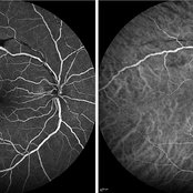
Fluorescein and Indocyanine Green Angiography of a 49-year-old male with high ANA titer, atypical ANCA positivity, who presented to clinic with 1 month of vision loss. Exam revealed anterior chamber cell, mild vitreous cell, sclerotic vessels along arterioles. Early FA/ICG at 1 minute demonstrates absent arteriole fill.
Photographer: Killian Roberts, Micaela Hertz; Eye Clinic of Wisconsin
Imaging device: Heidelberg Spectralis
Condition/keywords: A-ANCA, autoimmune vasculitis, fluorescein angiogram (FA), indocyanine green (ICG) angiography, retinal vasculitis
-
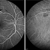 Late FA/ICG at 4 Minutes of Atypical ANCA Associated Retinal Vasculitis
Nov 13 2024 by Deepak Sambhara, MD
Fluorescein and Indocyanine Green Angiography of a 49-year-old male with high ANA titer, atypical ANCA positivity, who presented to clinic with 1 month of vision loss. Exam revealed anterior chamber cell, mild vitreous cell, sclerotic vessels along arterioles. Late FA/ICG at 4 minutes demonstrates absent arteriole fill with venular periphlebitis.
Photographer: Killian Roberts, Micaela Hertz; Eye Clinic of Wisconsin
Imaging device: Heidelberg Spectralis
Condition/keywords: A-ANCA, autoimmune vasculitis, fluorescein angiogram (FA), indocyanine green (ICG) angiography, retinal vasculitis
-
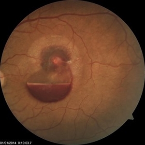
 Valsalva Retinopathy
Oct 28 2024 by Andrew Jin, MD
These fundus photos show the progression of subhyaloid hemorrhage due to valsalva retinopathy in a 38-year-old woman with hyperemesis gravidarum. She was managed conservatively with observation alone. The photos depict the initial presentation after 1 day of vision loss, at 1 month, and at 6 months of follow-up. Presenting visual acuity was counting fingers and returned to 20/20.
Condition/keywords: fundus photograph, valsalva retinopathy
-
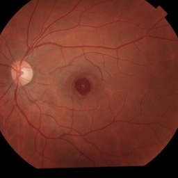
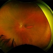
 Retinitis Pigmentosa
Oct 16 2024 by Virginia Gebhart
74 year old female with bone spicule pigmentation associated with Retinitis Pigmentosa. Pt diagnosed at age 53, relatively asymptomatic prior to diagnosis. Pt reports gradual vision loss over 10+ years. BCVA 20/40
Photographer: Virginia Gebhart, Retina Consultants of Carolina
Imaging device: Optos California
Condition/keywords: bone spicule, retinitis pigmentosa, retinitis pigmentosa (RP) dystrophy
-
 A Fleet of Boat-Shaped Hemorrhages
Aug 1 2024 by James P Dossett, MD
Pseudocolor fundus photograph of the left eye of a 54-year-old diabetic man presenting with bilateral vision loss. Examination revealed 20/200 vision OS with extensive preretinal and vitreous hemorrhage, marked diffuse neovascularization, macular edema and hard exudates.
Photographer: Beth Smith, West Virginia University Eye Institute
Condition/keywords: proliferative diabetic retinopathy (PDR)
-
 Choroidal Osteoma With CNV- Aautofluorescence
Feb 28 2024 by stephen oconnell
23 year old male with 10 months of vision loss prior to presentation. Clarus 700 autofluorescence image.
Condition/keywords: macular choroidal osteoma
 Loading…