Search results (19 results)
-
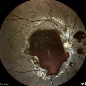
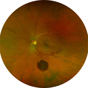 Large Subhyaloid Hemorrhage
Jul 11 2025 by Jessilla Phou
This is a fundus photograph depicting a large subhyaloid hemorrhage in the mid periphery of the left eye. The patient, a 53-year-old female, presented with a sudden onset of floaters, headache, and blurred vision. The image also demonstrates associated optic disc hemorrhage, vitreous hemorrhage, retinal hemorrhage, and venous tortuosity. Despite the extensive workup performed and the severity of the hemorrhage, no underlying cause was determined.
Photographer: Jessilla Phou
Imaging device: Optos California
Condition/keywords: fundus photograph, optic disc hemorrhage, retinal hemorrhage, venous tortuosity, vitreous hemorrhage
-
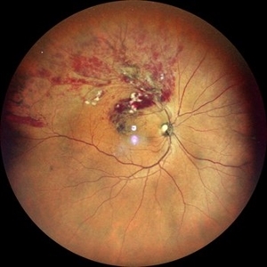 Branch Retinal Vein Occlusion
Apr 28 2024 by Anjana Mirajkar, MS Ophthalmology
A widefield color photo of a 55 year old male case of supero-temporal BRVO showing venous tortuosity, cotton wool spots, flame shaped hemorrhages and macular edema.
Photographer: Dr. Anjana Mirajkar -Retina Foundation, Ahmedabad
Imaging device: Mirante-Nidek
Condition/keywords: branch retinal vein occlusion (BRVO), ST BRVO
-
 Central Retinal Vein Occlusion (CRVO)
Sep 14 2023 by Ben Serar
Fundus photograph showing extensive scattered superficial retinal haemorrhages with retinal venous tortuosity and engorgement , along with disc edema in a case of Central Retinal Vein Occlusion (CRVO).
Condition/keywords: central retinal vein occlusion (CRVO), disc edema, Engorged veins, Tortuous veins
-
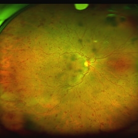
 Anaemic Retinopathy
Sep 13 2023 by Anand Temkar
Wide field image of the RE of a 35 year old male patient showing Roth's spots in all four quadrants and venous tortuosity in a case of Anaemic Retinopathy.
Photographer: Dr.Anand Temkar- Retina Foundation, Ahmedabad
Imaging device: Mirante
Condition/keywords: anaemic retinopathy, roth spots
-
 Anemic Retinopathy Related Retinal Hemorrhages
Nov 5 2019 by Chinmayi Vyas
Anemic retinopathy related retinal hemorrhages in a 24 years old male with Hb of 4.2gm/ dl. The manifestations of anemic retinopathy are nonspecific and may closely simulate hypertensive or diabetic retina. Retinal changes in anemia are cotton wool spots, venous tortuosity, and hemorrhages which may be present at all levels of the retina and choroid. All retinal hemorrhages can occur when Hb falls below 8 g/100 ml or if the platelet count falls below 50,000/cumm. The combination of severe anemia and thrombocytopenia is likely to produce retinal hemorrhages. The Roth’s spots or white centre hemorrhages are typically associated with bacterial endocarditis , anemia and other systemic conditions. The white center is suspected to represents focal ischemia, inflammatory or infectious infiltrate, fibrin or accumulation of neoplasticism cells.
Photographer: Dr Chinmayi Vyas, Nethradhama superspeciality eye hospital , banglore, india
Imaging device: Eidon fundus imaging
Condition/keywords: anaemic retinopathy
-
 Anemic Retinopathy Related Retinal Hemorrhages
Nov 5 2019 by Chinmayi Vyas
Anemic retinopathy related retinal hemorrhages in a 24 years old male with Hb of 4.2gm/ dl. The manifestations of anemic retinopathy are nonspecific and may closely simulate hypertensive or diabetic retina. Retinal changes in anemia are cotton wool spots, venous tortuosity, and hemorrhages which may be present at all levels of the retina and choroid. All retinal hemorrhages can occur when Hb falls below 8 g/100 ml or if the platelet count falls below 50,000/cumm. The combination of severe anemia and thrombocytopenia is likely to produce retinal hemorrhages. The Roth’s spots or white centre hemorrhages are typically associated with bacterial endocarditis , anemia and other systemic conditions. The white center is suspected to represents focal ischemia, inflammatory or infectious infiltrate, fibrin or accumulation of neoplasticism cells.
Photographer: Dr Chinmayi Vyas
Condition/keywords: retinal hemorrhage
-
 Central Retinal Vein Occlusion
Dec 22 2018 by FELIPE PEREIRA
25-year-old male patient with acute and painless vision loss of left eye. The fundus examination demonstrate optic disc swelling, venous tortuosity, diffuse intraretinal hemorrhage and severe macular edema. There is also extensive exudative retinal detachment with lipid deposits in the posterior pole, mainly around the vessels. The systemic work up was negative, including serologies, rheumatologic and hematological markers and cholesterol and triglycerides within normal limits.
Photographer: Felipe Pereira
Imaging device: Vizucan, Zeiss
Condition/keywords: central vein occlusion, ischemic CRVO
-
 Proliferative Sickle Cell Retinopathy, Red Free OS
May 23 2018 by Hosam Attia, MD
Red free fundus photograph of a 45-year-old African American, male with sickle cell anemia (SC disease) with arteriolar attenuation, mild venous tortuosity, peripheral arterio-venous anastomoses (Inferotemporally), multiple small NVEs/ early sea fans OS.
Photographer: Aaron Appiah, M.D.
Imaging device: Optos California Ultra-Wide Field Fundus Camera
Condition/keywords: neovascularization elsewhere (NVE), proliferative retinopathy, sea fan, sickle cell, sickle cell retinopathy
-
 Proliferative Sickle Cell Retinopathy, Red Free OD
May 23 2018 by Hosam Attia, MD
Red free fundus photo of a 45-year-old African American, male with sickle cell anemia (SC Disease ) with arteriolar attenuation, mild venous tortuosity, Sunburst (S) and large, partially auto-infarcted sea fan, OD.
Imaging device: Optos California Ultra-Wide Field Fundus Camera
Condition/keywords: neovascularization elsewhere (NVE), proliferative retinopathy, sea fan, sickle cell, sickle cell retinopathy
-
 Proliferative Sickle Cell Retinopathy, Color OS
May 23 2018 by Hosam Attia, MD
45-year-old African American, male with sickle cell anemia (SC disease ) with arteriolar attenuation, mild venous tortuosity, peripheral arterio-venous anastomoses (shown better on red free), multiple small NVEs/ early sea fans (one w/ early auto-infarction) and sunburst (S) - (Not showing very well in photos) OS.
Imaging device: Optos California Ultra-Wide Field Fundus Camera
Condition/keywords: neovascularization elsewhere (NVE), proliferative retinopathy, sea fan, sickle cell, sickle cell retinopathy
-
 Proliferative Sickle Cell Retinopathy, Color OD
May 23 2018 by Hosam Attia, MD
45-year-old African American, male with sickle cell anemia (SC disease) with arteriolar attenuation, mild venous tortuosity, Sunburst (S) and large, partially auto-infarcted Seafan with fresh heme, OD.
Imaging device: Optos California Ultra-Wide Field Fundus Camera
Condition/keywords: neovascularization elsewhere (NVE), proliferative retinopathy, sea fan, sickle cell, sickle cell retinopathy
-
 Proliferative Sickle Cell Retinopathy, Color OD
May 23 2018 by Hosam Attia, MD
45-year-old African American, male with sickle cell anemia (SC disease) with arteriolar attenuation, mild venous tortuosity, Sunburst (S) and large, partially auto-infarcted sea fan with fresh heme, OD.
Imaging device: Optos California Ultra-Wide Field Fundus Camera
Condition/keywords: neovascularization elsewhere (NVE), proliferative retinopathy, sea fan, sickle cell, sickle cell retinopathy
-
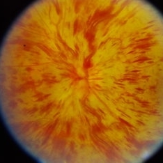
 Idiopathic Papillophlebitis- Fluorescein Angiography
Apr 5 2017 by Linda A Cernichiaro- Espinosa, MD
18-year-old otherwise healthy female with sudden visual loss on the left eye.
Photographer: Linda A Cernichiaro MD
Imaging device: Optos Daytona
Condition/keywords: cystoid macular edema (CME), papilledema, venous tortuosity
-
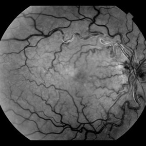
 CRVO
CRVO
Sep 7 2015 by Andrea Arriola-Lopez, MD MSc
Color fundus photography of right eye with central retinal vein occlusion; BCVA 20/25; IOP 16mmHg. No macular edema or neovascularization.
Photographer: Andrea Elizabeth Arriola López, MD, MSc
Imaging device: OPTOS Dakota
Condition/keywords: blot hemorrhages, central retinal vein occlusion (CRVO), venous tortuosity
-
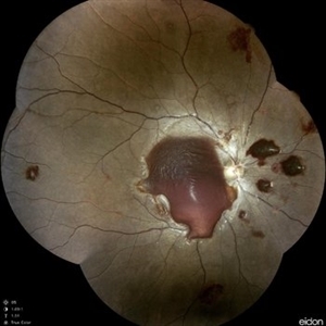
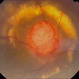
 Red Free Photo of Optic Disc Capillary Hemangioblastoma
Mar 18 2014 by Arwa Azmeh, MD, PhD
Red free fundus photograph of an 48-year-old male who complained of decreased visual acuity in his right eye over the last few months. Systemically the patient was healthy. His VA was OD Cf 3m, OS 20/20. Anterior segments were WNL in OU. IOP was WNL in OU. Fundus exam OD revealed unpigmented mass over the optic disc with retinal venous tortuosity at its edges with a ring of thick HYE surrounding it and shallow RD in this area extending to the foveal area. Several few small retinal hemorrhages were seen in the far retinal periphery which were explained to be caused by venous stasis due to the optic disc tumor
Condition/keywords: optic disc, red-free, retinal hemangioblastoma
-
 Color Photo of Optic Disc Capillary Hemangioblastoma
Mar 18 2014 by Arwa Azmeh, MD, PhD
Color fundus photograph of an 48-year-old male who complained of decreased visual acuity in his right eye over the last few months. Systemically the patient was healthy. His VA was OD Cf 3m, OS 20/20. Anterior segments were WNL in OU. IOP was WNL in OU. Fundus exam OD revealed unpigmented mass over the optic disc with retinal venous tortuosity at its edges with a ring of thick HYE surrounding it and shallow RD in this area extending to the foveal area. Several few small retinal hemorrhages were seen in the far retinal periphery which were explained to be caused by venous stasis due the optic disc tumor.
Condition/keywords: color photo, optic disc, retinal hemangioblastoma
-
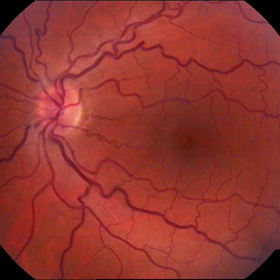 Congénital Venous Tortuosity OS
Mar 13 2013 by Jose Dalma-Weiszhausz, MD
Young patient with routine normal eye exam.
Photographer: José Dalma, MD Dalma & Assoc., Mexico City
Imaging device: Topcon 50VT
Condition/keywords: congenital venous tortuosity
-
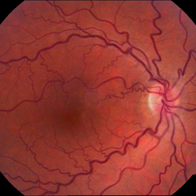 Congenital Venous Tortuosity OD
Mar 13 2013 by Jose Dalma-Weiszhausz, MD
Venous tortuosity in a young patient in for routine eye exam.
Photographer: José Dalma, MD Dalma & Assoc., Mexico City
Condition/keywords: congenital venous tortuosity
-
 Central Retinal Vein Occlusion
Oct 13 2012 by Geoffrey G. Emerson, MD, PhD, FASRS
Central retinal vein occlusion
Condition/keywords: central retinal vein occlusion (CRVO), venous tortuosity
 Loading…