Search results (13 results)
-
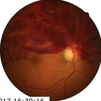
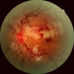
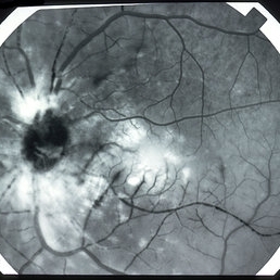
 Branch Retinal Vein Occlusion
Apr 15 2023 by Yousef A Fouad, MBBCh, MSc
BRVO in a young female with uncontrolled hypertension
Photographer: Yousef Fouad, Ain Shams University, Egypt
Condition/keywords: branch retinal vein occlusion (BRVO), fundus photograph, Hypertension, venous occlusion
-
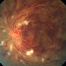
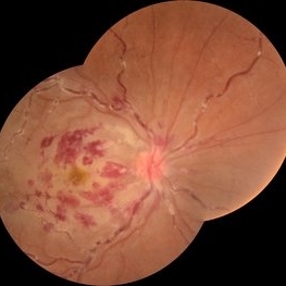 Combined Central Retinal Artery Occlusion with Central Retinal Venous Occlusion
Mar 22 2023 by VIRAL SHAH
26 YEARS OLD MALE PATIENTS HAS COMPLAIN OF DIMNESS OF VISION SINCE 3 DAYS IN RIGHT EYE. HE IS SUFFERING FROM ANEMIA
Photographer: VIRAL SHAH
Condition/keywords: VASCULAR SHEATHING WITH HOLLENHORST PLAQUE
-
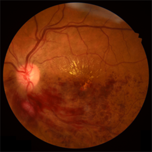
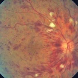
 Combined central retinal vein occlusion and branch retinal arteriolar occlusion
Sep 13 2022 by Ruchir Mehta, DO, DNB, FRCS
Fundus photograph of left eye of a 63 years old female with known type 2 DM and HTN showing combined central retinal venous occlusion and superior branch retinal arteriolar occlusion
Photographer: Ruchir Mehta, Mehta Superspeciality Eye Hospital, Jamnagar, Gujarat, India
Imaging device: Zeiss Visucam 500
Condition/keywords: branch retinal artery occlusion (BRAO), central retinal vein occlusion (CRVO), COMBINED
-
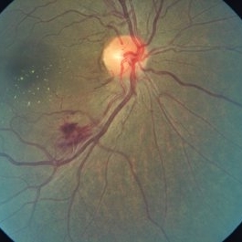
 Retinal Macroaneurysm
Dec 17 2019 by Jonathan C. Tsui, MD
A 72-year-old female with uncontrolled hypertension presented with several spots in her vision. Fundus photography demonstrated a retinal macroaneurysm hemorrhage with subretinal fluid and intraretinal heme. Several microaneurysms are present adjacent to an anomalous vein which suggests a possible secondary venous occlusion.
Condition/keywords: retinal macroaneurysm
-
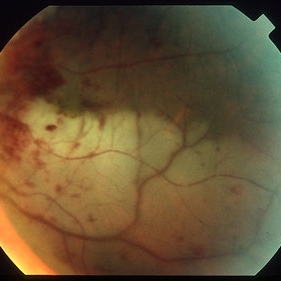
 Branch Venous Occlusion
Oct 20 2018 by Jose Lopez
Branch venous occlusion.
Photographer: Irene López Liroz
Condition/keywords: branch retinal vein occlusion (BRVO)
-
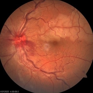
 Hemi-Central Retinal Venous Occlusion
Apr 17 2018 by Ronald Silva
Fundus photograph of an 55-year-old man with low vision acuity for 2 weeks, and was observed hemi-central retinal venous oclusion right eye.
Photographer: Ronald Rocha da Silva, HCOE, Feira de Santana-BA
Condition/keywords: central retinal vein occlusion (CRVO)
-
 Combined Cilioretinal Artery and Central Retinal Vein Occlusion
May 14 2016 by Ines Leal
Combined cilioretinal artery and central retinal vein occlusion in an otherwise 49-year-old healthy female patient. Color fundus photography shows whitening of the retina in the distribution of the cilioretinal artery and intraretinal hemorrhages with tortuous and engorged veins.
Photographer: Inês Leal, MD, Department of Ophthalmology, Faculty of Medicine, Universidade de Lisboa,
Condition/keywords: cilioretinal artery occlusion, venous occlusion
-
 Central Retinal Vein Occlusion
Apr 24 2015 by Mehul A Shah
A 55-year-old female presented with sudden loss of vision, she is known case of systemic hypertension presenting vision was FCNF and had severe macular edema.
Photographer: Mehul Shah, Drashti Netralaya
Imaging device: Zeiss FF450plus
Condition/keywords: central retinal vein occlusion (CRVO), venous occlusion
-
 Central Retinal Vein Occlusion
Apr 23 2015 by Mehul A Shah
Patient presented with sudden loss of vision and patient had systemic hypertension.
Photographer: Mehul Shah
Imaging device: Zeiss FF450 Plus
Condition/keywords: central retinal vein occlusion (CRVO), venous occlusion
-
 Impending Branch Retinal Vein Occlusion
Apr 23 2015 by Mehul A Shah
Patient presented with diminished vision found to have vision 6/12 and patient became 6/6 after anti vegf injection.
Photographer: Mehul Shah
Imaging device: Zeiss FF450 Plus
Condition/keywords: branch vein occlusion (BVO), venous occlusion
-
 Behcet's FA 2
Jan 11 2013 by Alex P. Hunyor, MD
Behcet's disease - combined arterial / venous occlusion. Fluorescein angiogram 2 of 2, left eye.
-
 Behcet's FA 1
Jan 11 2013 by Alex P. Hunyor, MD
Behcet's disease - combined arterial / venous occlusion. Fluorescein angiogram 1 of 2, left eye.
-
 Behcets colour
Jan 11 2013 by Alex P. Hunyor, MD
Behcet's disease - combined arterial / venous occlusion. Color image, left eye.
 Loading…