Search results (43 results)
-
 Retinocoroiditis Inactiva Por Toxoplasmosis
Apr 28 2025 by Paulina Araujo
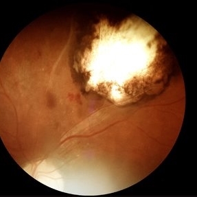
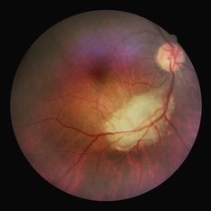
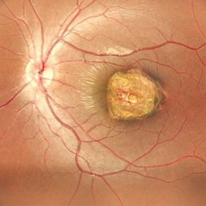
Fundus photography demonstrates a 2-disc-diameter chorioretinal scar in the superior temporal arcade, consistent with inactive toxoplasmic retinochoroiditis. The lesion exhibits pigmented borders and central atrophy, with adjacent splinter hemorrhages and vascular sheathing. No vitreous inflammation or active satellite lesions are present.
Photographer: Paulina D.Araujo Martínez, Asociación para Evitar la Ceguera en México I.A.P., Hospital Dr Luis Sánchez Bulnes.
Condition/keywords: toxoplasmosis chorioretinitis
-
 Toxoplasmosis Disease
Mar 30 2024 by Karen Flores Guevara
Fundus photograph of a 7-year-old-child with a macular scar observed over time for growth.
Photographer: Diana Elizabeth Arellano Acosta MD Pediatric Retina,Asociación para Evitar la Ceguera en México IAP. México
Condition/keywords: toxoplasmosis chorioretinitis
-
 Toxoplasmosis Chorioretinitis
Mar 2 2024 by James P Dossett, MD
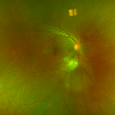
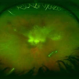
Pseudocolor fundus photograph of the right eye of a 34-year-old man with retinitis along the inferotemporal arcade with associated subretinal fluid and overlying vitritis. Aqueous paracentesis was performed and PCR was positive for Toxoplasma gondii. He was administered intravitreal clindamycin.
Imaging device: Optos
Condition/keywords: posterior uveitis, toxoplasmosis chorioretinitis
-
 Hidden Mickey / Toxo Scar
Feb 29 2024 by Virginia Gebhart
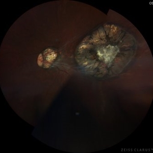
87 year old female with inactive toxoplasmosis chorioretinitis inferior. Stable regressed malignant neoplasm of choroid superior (s/p brachytherapy 2011).
Photographer: Virginia Gebhart
Imaging device: Optos California
Condition/keywords: inactive toxoplasmosis, toxo chorioretinitis, toxoplasmosis chorioretinitis
-
 Toxoplasmosis
Jul 14 2023 by Aditya Rali, MD
Toxoplasmosis chorioretinitis
Condition/keywords: ocular toxoplasmosis
-
 Ocular toxoplasmosis
Mar 5 2023 by Sergio Emilio Sifuentes Renteria, MD
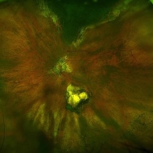
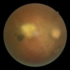
Color fundus photograph of the right eye of a patient with HIV-infection and concomitant ocular toxoplasmosis.
Photographer: Sergio Emilio Sifuentes Rentería - Clínica Especializada Condesa Iztapalapa
Condition/keywords: HIV, infectious uveitis, posterior uveitis, toxoplasmosis, toxoplasmosis chorioretinitis
-
 Choroidal neovascular membrane
Nov 4 2022 by rodrigo torres
Neovascular membrane on edge of toxoplasmosis chorioretinitis scar.
Photographer: Rodrigo Torres
Condition/keywords: choroidal neovascular membrane (CNVM), toxoplasmosis chorioretinitis, uveitis
-
 Toxoplasmic chorioretinitis
Nov 4 2022 by rodrigo torres
Fundus photograph of an 20 yrs old woman whit active lesion of toxoplasmosis chorioretinitis. Hiperpigmented scar associated in macular region
Photographer: Rodrigo Torres
Condition/keywords: toxoplasmosis chorioretinitis, uveitis
-
 Ocular Toxoplasmosis
May 6 2022 by Eder Díaz Dorado
Fundus photograph of an 31-year-old man with a macular scar of toxoplasmosis and a review with OCT
Photographer: Eder Díaz Dorado, Hospital Central Militar, Ciudad de México
Imaging device: Heidelberg Spectralis /Smartphone Photography
Condition/keywords: inactive toxoplasmosis, ocular toxoplasmosis, toxoplasmosis chorioretinitis
-
 Recurrence of Ocular Toxoplasmosis
Apr 11 2022 by Aniruddha K Agarwal, MD
Inactive toxoplasmosis lesion with active lesion at the inferior edge. The active lesion appears "fuzzy". Note also the overlying vitritis
Photographer: Debra A. Goldstein, MD, FRCSC Magerstadt Professor of Ophthalmology Director, Uveitis Service Director, Uveitis Fellowship Department of Ophthalmology Northwestern University Feinberg School of Medicine
Condition/keywords: IUSG, recurrence, retinochoroiditis, toxoplasmosis chorioretinitis, uveitis
-
 Toxoplasmic Retinitis
Feb 11 2021 by David L Kilpatrick, MD
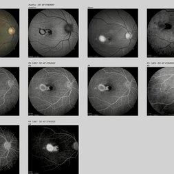
A 50-year-old female presented with a history of subacute central vision loss involving the right eye. Exam showed a focal, creamy white retinitis involving the fovea with mild vitritis. FA demonstrates blockage of the lesion initially followed by progressive leakage, most prominently at the border of the retinitis. Toxoplasma IgM and IgG were both markedly elevated. The patient was treated with a combination of Bactrim, Intravitreal Clindamycin, and Oral steroids.
Photographer: MS Retina Associoates
Imaging device: Optos
Condition/keywords: toxoplasmosis chorioretinitis, toxoplasmosis retinitis
-
 Toxoplasmic Chorioretinitis
May 18 2020 by McGill University Health Centre
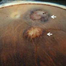
Toxoplasmic chorioretinitis is caused by parasitic infection from Toxoplasma gondii. Two forms are recognized: congenital and acquired. Congenital toxoplasmic chorioretinitis occurs because the infection is transplacental: T. gondii is among infections that cause TORCH syndrome. Acquired toxoplasmic chorioretinitis is produced by parasite ingestion, usually from raw or undercooked food. After parasitemia, the parasite directly invades the photoreceptors in the retina. In this enucleation specimen, chronic and subacute lesions coexist. In the active lesion located in the macula (arrow), the retina is necrotic, and reactive RPE cell hyperplasia surrounds the lesion. The chronic lesion (arrowhead) demonstrates atrophy of the retina and RPE in the center; in the periphery, RPE proliferation is present.
Condition/keywords: toxoplasmosis chorioretinitis
-
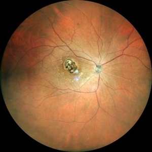
 Toxoplasma Scar [Color Photo]
Nov 15 2019 by Sham Talati, DOMS
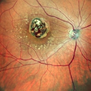
Fundus photograph of toxo scar on macula.
Photographer: Dr. Sham Talati,Retina Foundation,Ahmedabad
Imaging device: Nidek Mirante
Condition/keywords: toxoplasmosis, toxoplasmosis chorioretinitis
-
 Toxoplasma Scar [Color Photo]
Nov 15 2019 by Sham Talati, DOMS
Fundus photograph of toxo scar on macula.
Photographer: Dr. Sham Talati,Retina Foundation,Ahmedabad
Imaging device: Nidek Mirante
Condition/keywords: toxoplasmosis, toxoplasmosis chorioretinitis
-
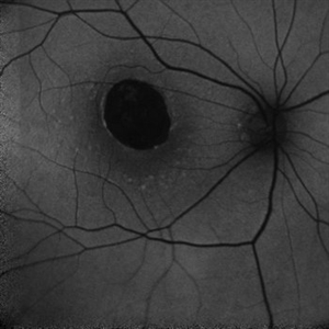
 Toxoplasma Scar [Retro mode]
Nov 15 2019 by Sham Talati, DOMS
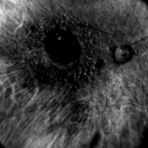
Fundus photograph of toxo scar on macula.
Photographer: Dr. Sham Talati,Retina Foundation,Ahmedabad
Imaging device: Nidek Mirante
Condition/keywords: toxoplasmosis, toxoplasmosis chorioretinitis
-
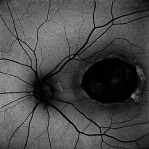
 Toxoplasma Scar [Retro mode]
Nov 15 2019 by Sham Talati, DOMS
Fundus photograph of toxo scar on macula.
Photographer: Dr. Sham Talati,Retina Foundation,Ahmedabad
Imaging device: Nidek Mirante
Condition/keywords: toxoplasmosis, toxoplasmosis chorioretinitis
-
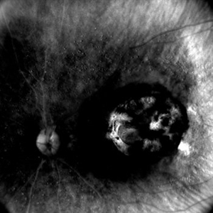
 Toxoplasma Scar [Autofluorescence]
Nov 15 2019 by Sham Talati, DOMS
Fundus photograph of toxo scar on macula.
Photographer: Dr. Sham Talati,Retina Foundation,Ahmedabad
Imaging device: Nidek Mirante
Condition/keywords: toxoplasmosis, toxoplasmosis chorioretinitis
-
 Toxoplasma Scar [Autofluorescence]
Nov 15 2019 by Sham Talati, DOMS
Fundus photograph of toxo scar on macula.
Photographer: Dr. Sham Talati,Retina Foundation,Ahmedabad
Imaging device: Nidek Mirante
Condition/keywords: toxoplasmosis, toxoplasmosis chorioretinitis
-
 Toxoplasma Scar [Color Photo-Wide Field]
Nov 15 2019 by Sham Talati, DOMS
Fundus photograph of toxo scar on macula.
Photographer: Dr. Sham Talati,Retina Foundation,Ahmedabad
Imaging device: Nidek Mirante
Condition/keywords: toxoplasmosis, toxoplasmosis chorioretinitis
-
 Toxoplasma Scar [Color Photo-Wide Field]
Nov 15 2019 by Sham Talati, DOMS
Fundus photograph of toxo scar on macula.
Photographer: Dr. Sham Talati,Retina Foundation,Ahmedabad
Imaging device: Nidek Mirante
Condition/keywords: toxoplasmosis, toxoplasmosis chorioretinitis
-
 Toxo Lesion on Macula
Jul 25 2019 by Manish Nagpal, MD, FRCS (UK), FASRS
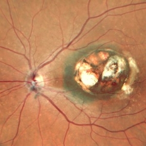
Fundus photo of old toxo lesion on macula.
Photographer: Gayathri Mohan, Retina Foundation
Imaging device: Nidek Mirante SLO
Condition/keywords: toxoplasmosis, toxoplasmosis chorioretinitis
-
 Recurrent Toxoplasmosis
Apr 8 2019 by Gary R. Cook, MD, FACS
17-year-old male with recurrent toxoplasmosis lesion adjacent to an earlier, inactive toxoplasmosis chorioretinal scar superior to the optic disc OS. The recurrent lesion is becoming less inflamed and 'harder' in appearance, indicating resolution
Condition/keywords: toxo chorioretinitis, toxoplasmosis, toxoplasmosis chorioretinitis, toxoplasmosis reactivation
-
 Toxoplasma Scar
Sep 22 2018 by Hashim Ali Khan, OD, FAAO
Fundus photograph of a 17-year-old male with inactive macular toxoplasma scar.
Condition/keywords: inactive toxoplasmosis, toxoplasmosis chorioretinitis
-
 Toxoplasma Macular Scar
Sep 22 2018 by Hashim Ali Khan, OD, FAAO
Fundus Photographs of a 17-year-old male with inactive macular toxoplasma scar.
Condition/keywords: inactive toxoplasmosis, toxoplasmosis chorioretinitis
-
 Retinal Toxoplasmosis
Jul 20 2018 by Marco D'Angelo
Right eye, 50-year-old Caucasian woman, 14/20 visual acuity.
Photographer: Dott. Marco D'Angelo, S. Chiara Hospital, Trento, Italy
Imaging device: Topcon TRC-NW 6S
Condition/keywords: chorioretinal scar, toxoplasmosis chorioretinitis
 Loading…