Search results (73 results)
-
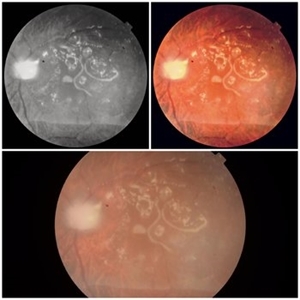
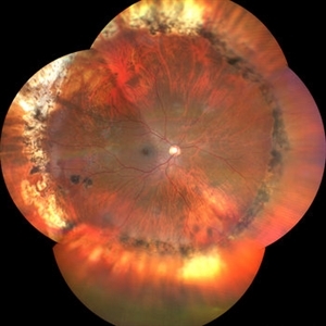
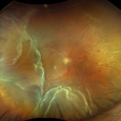 Total Retinal Detachment
Aug 6 2025 by Korey Starkey
59 year-old patient presents with total retinal detachment at first visit in OD. Recommending prompt surgical intervention.
Photographer: Korey Starkey
Imaging device: Optos
Condition/keywords: color fundus photograph, Optos, retinal detachment, total retinal detachment
-
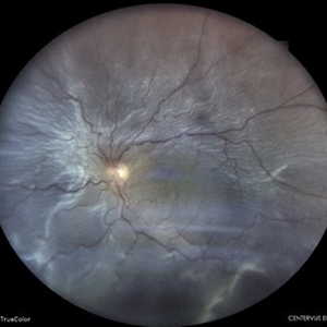
 Retinal Detachment
Aug 4 2025 by Anjana Mirajkar, MS Ophthalmology
Fundus photograph of a 40 year old male with a total retinal detachment with macula off with a HST at 1 o clock and a break at mid periphery at 8 o clock.
Photographer: Dr. Anjana Mirajkar- HV Desai eye hospital ,Pune
Imaging device: optos
Condition/keywords: retinal detachment, retinal holes
-
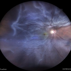
 Rhegmatogenous Retinal Detachment with Gd C PVR Changes
Mar 28 2025 by Shrishti mishra
Fundus photograph of a 58 year old male who had undergone a pneumatic retinopexy elsewhere presented to us with a total retinal detachment with a retinal tear in the superotemporal quadrant and grade c pvr changes.
Photographer: Mrs Vinutha
Imaging device: Optos nikon
Condition/keywords: proliferative vitreoretinopathy (PVR), retinal tear with detachment, rhegmatogenous retinal detachment
-
 Repaired Retinal Detachment with Grade C PVR
Dec 23 2024 by Virginia Gebhart
61 year old male 1 day s/p retinectomy/SO exchange. Retina is attached under SO with good laser to retinectomy edge.
Photographer: Virginia Gebhart, Retina Consultants of Carolina
Imaging device: Optos California
Condition/keywords: gas bubble, proliferative vitreoretinopathy (PVR), retinectomy, silicone oil, total retinal detachment
-
 Morning Glory Anomaly With Retinal Detachment Managed With Amniotic Membrane Graft
Oct 15 2024 by Hemanth Murthy, MBBS, MD, FASRS
10 year-old boy presented with noticed blurring of vision. He had total retinal detachment with complicated cataract. He underwent lensectomy with 240 band and vitrectomy with silicone oil. The retina failed to settle due to minute breaks in the inferior part of the disc. Repeat surgery with AMG was done to cover the inferior part of disc. The retina settled under silicone oil. Silicone oil was removed and he is presently undergoing amblyopia treatment. Vision is 2/60 with +14.5 diopter lens.
Photographer: Mr Veda Vyas
Condition/keywords: amniotic membrane graft, Morning Glory Anomaly
-
 Emulsified Silicone Oil in Macular Hole
Jun 7 2024 by Vaidehi Jethwa
Fundus photograph of 72 year old man was having a/h/o Left Eye trauma by a cow horn, 8 years Ago, and developed Left Eye total Retinal detachment and was operated for Left Eye vitrectomy with FAX, SOI, Endolaser on 11 /04/2015 and was advised Left Eye silicone Oil removal.
Photographer: Dr. Vaidehi Jethwa, M and J institute of Ophthalmology, Ahmedabad, Gujarat.
Imaging device: Zeiss Visucam lite
Condition/keywords: macular hole, silicon oil emulsification in vitreous cavity
-
 Retinal Detachment Status Post Trauma
May 7 2024 by Akansha Sharma
Color fundus photograph of a 47 year old male with total retinal detachment.
Photographer: Dr. Akansha Sharma, Bharati Eye Hospital
Condition/keywords: RD, Retinal Detachment
-
 Giant Retinal Tear
Feb 20 2024 by Soobien Lee
Optos color fundus photograph of a 40-year-old caucasian male who is a UFC fighter with a total retinal detachment in his right eye secondary to a giant retinal tear from 10 o'clock to 2 o'clock.
Photographer: Trinity Wolf, Elman Retina Group
Imaging device: Optos Ultra-Widefield Imaging
Condition/keywords: giant retinal tear, optos, Retinal Detachment, Retinal tear with detachment, trauma
-
 Sub-total Retinal Detachment
Jan 30 2024 by Akansha Sharma
Color fundus photograph of a 53 year old male patient with sub-total retinal detachment.
Photographer: Dr. Akansha Sharma, Bharati Eye Hospital
Condition/keywords: RD, Retinal Detachment, sub-total retinal detachment
-
 Chronic Retinal Detachment with Proliferative Vitreoretinopathy
Jan 25 2024 by Isaac Agranoff
Widefield fundus photography of a 24 year old male presenting with subtotal retinal detachment with circumferential anterior proliferative vitreoretinopathy. The detachment is bullous inferiorly with atrophic retina and subretinal bands. There are also scattered patches of lattice with atrophic holes and associated detachment in the periphery. Patient presented with flashes for 2 years with worsening vision over the past 6-8 months, measured at 20/100 ph 20/60 OS.
Photographer: Isaac Agranoff, Ashley Rigdon
Imaging device: Optos California
Condition/keywords: atrophic hole, chronic retinal detachment, lattice degeneration, proliferative vitreoretinopathy (PVR), subretinal bands
-
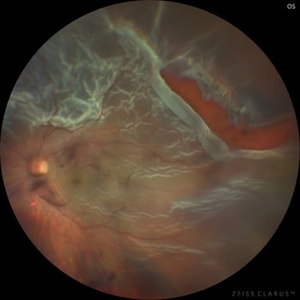 Total Rhegmatogenous retinal detachment with lattice degeneration & Vitreous haemorrhage
Jul 31 2023 by Harsh Vardhan Singh, MS
72-year male presented PVD induced total retinal detachment with vitreous hemorrhage
Photographer: Dr Harsh Vardhan Singh, AIIMS, Guwahati
Imaging device: Zeiss Clarus 700
Condition/keywords: chronic retinal detachment, hemorrhage, rrd
-
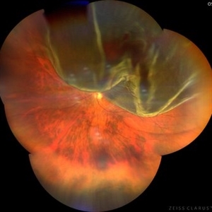
 Total Rhegmatogenous retinal detachment with opened posterior margin of lattice degeneration
Jul 18 2023 by Harsh Vardhan Singh, MS
78-year-old man with history of defective following cataract surgery showed total retinal detachment on examination
Photographer: Harsh Vardhan Singh, AIIMS, Guwahati
Imaging device: Zeiss Clarus 700
Condition/keywords: chronic retinal detachment, peripheral lattice degeneration, rrd
-
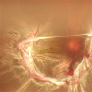
 Superior bullous retinal detachment
Jan 14 2023 by Vishal Agrawal, MD, FRCS,FACS,FASRS
50 year old female with left eye sub total retinal detachment
Photographer: Pankaj
-
 Stickler syndrome with retinal detachment
Jan 14 2023 by Vishal Agrawal, MD, FRCS,FACS,FASRS
30 year old female with stickler syndrome and subtotal retinal detachment . Fundus pic shows radial para vascular lattices with multiple breaks .
Photographer: Pankaj
Imaging device: CLARUS 700
Condition/keywords: Retinal Detachment, Stickler Syndrome
-
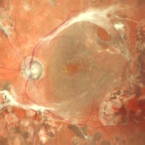
 Post PPV for retinal detachment
Oct 13 2022 by Vishal Agrawal, MD, FRCS,FACS,FASRS
3 month Post operative picture of right eye .The patient had PPV + gas (C3F8) for total retinal detachment with multiple breaks.
Photographer: Pankaj, Agrawal Hospital,Jaipur
Imaging device: Clarus 700
-
 Total retinal Detachment multiple holes
Sep 26 2022 by Denica Rodriguez
60 year old Male presented with two week old Macula off Retinal detachment with multiple tears.
Photographer: Denica Rodriguez
Imaging device: Optos California
Condition/keywords: color fundus photograph, color photo, macula-off, optos, pseudocolor, Retinal detachment, retinal holes, retinal tear, Retinal tear with detachment, superior arcade, superior field, superior retina, total retinal detachment
-
 Retinal Detachment
Sep 9 2022 by Vishal Agrawal, MD, FRCS,FACS,FASRS
24-year-old female patient presented with sudden decrease in vision. On examination there was a left eye subtotal retinal detachment involving macula.
Photographer: Vishal Agrawal MD
Imaging device: Clarus 700
Condition/keywords: bullous retinal detachment, retinal break
-
 ROP 5A
Jan 24 2022 by Alexandre Grandinetti, MD, PhD
ROP retinal detachment.
Photographer: Alexandre Grandinetti
Imaging device: RetCam
Condition/keywords: retinopathy of prematurity (ROP), total retinal detachment
-
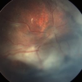
 Retinal Detachment post ROP
Jul 22 2021 by Vishal Gupta, MBBS, MS
Fundus image of total Retinal Detachment in a five-year-old male kid with a history of prematurity.
Photographer: Dr Shobhit Chawla, Prakash Netra Kendr, Lucknow, UP, INDIA
Imaging device: Zeiss Clarus
Condition/keywords: retinopathy of prematurity (ROP)
-
 RD Montage
Jul 3 2021 by Somnath Chakraborty, MD
Fundus photo montage of the left eye of a 56-year-old male showing subtotal retinal detachment with macular involvement and a large circumlinear tear extending from 1 o' clock to 3 o' clock hours.
Photographer: Pulak Roy
Condition/keywords: acute retinal detachment, retinal detachment of the macula, retinal tear, retinal tear with detachment
-
 Macrocyst in the Fovea
Feb 2 2021 by Peter J Belin, MD
36-year-old male with a white cataract and a chronic total retinal detachment for 1 year presented with a recurrent PVR detachment after primary repair 2 weeks prior. This OCT- EDI demonstrates a large retinal cyst through the fovea.
Photographer: Holly Cheshier, CRA, OCT-C, COT
Imaging device: Heidelberg Spectralis
Condition/keywords: chronic retinal detachment, proliferative vitreoretinopathy (PVR), retinal cyst, retinal macrocyst
-
 Retinal Detachment
Aug 23 2020 by Renata Bertazzi
Fundus photograph of an 76-year-old man with a total retinal detachment.
Photographer: Renata Bertazzi, Instituto Paulista de ensino e Pesquisa em Oftalmologia, São Paulo, SP
Imaging device: DayTona - Optos
Condition/keywords: acute retinal detachment
-
 TRD in Lasered PDR
Jun 10 2020 by Manish Nagpal, MD, FRCS (UK), FASRS
TRD in lasered PDR.
Photographer: gayathri mohan
Imaging device: nidek slo mirante
Condition/keywords: proliferative diabetic retinopathy (PDR), total retinal detachment
-
 Tractional retinal detachment
Jun 10 2020 by Manish Nagpal, MD, FRCS (UK), FASRS
TRD in PDR
Photographer: gayathri mohan
Imaging device: nidek slo mirante
Condition/keywords: proliferative diabetic retinopathy (PDR), total retinal detachment, tractional retinal detachment
 Loading…