Search results (274 results)
-
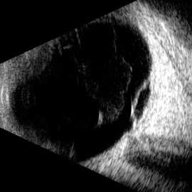
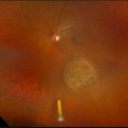
 New Collar Button Melanoma
Jan 7 2026 by Virginia Gebhart
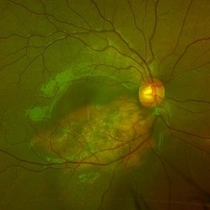
67 year old female referred for possible choroidal melanoma. Clinical exam, photos and ultrasound findings consistent with melanoma. Large pigmented tumor with collar button configuration, subretinal fluid inferior and vitreous hemorrhage. Left eye has scleral pigmentation 360 degrees, pt states she has had "brown spots" on her eye since childhood. Iris and fundus are also noticeably darker in the left eye consistent with oculodermal melanocytosis. Pt states she has had regular eye exams and has never been diagnosed. Pt will be scheduled for plaque brachytherapy pending CT scan results.
Photographer: Virginia Gebhart, Retina Consultants of Carolina
Imaging device: Optos California
Condition/keywords: choroidal melanoma, collar button, hemorrhage, melanoma, Oculodermal Melanocytosis
-
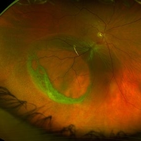
 Rhegmatogenous Retinal Detachment
Nov 27 2025 by Jacob Adrián Ruíz García
Ultra–widefield fundus image demonstrates an extensive rhegmatogenous retinal detachment (RRD) with several large, irregular retinal tears are visible in the temporal retina, with associated surrounding subretinal fluid. The detachment appears bullous, with fluid extending widely across the mid-periphery and involving much of the posterior pole.
Photographer: Jacob Adrián Ruíz Garcia, Fundación Hospital Nuestra Señora de la Luz I.A.P, México City
Imaging device: Optos California
Condition/keywords: horseshoe tear, Rhegmatogenous retinal detachment, tear
-
 Retinal Break With Cuff of Subretinal Fluid (Subclinical Retinal Detachment), Immediately Post Laser Barrage
Nov 26 2025 by NIDHI PANWAR, MD FRCS Glasgow FNB FICO
Immediately post laser barrage, case of asymptomatic retinal break with subretinal fluid more than 2 DD in supero temporal quadrant.
Photographer: NIDHI PANWAR
Imaging device: optos
Condition/keywords: asymptomatic retinal break, asymptomatic retinal detachment, BARRAGE LASER, subretinal fluid
-
 Retinal Break With Cuff of Subretinal Fluid (Subclinical Retinal Detachment)
Nov 26 2025 by NIDHI PANWAR, MD FRCS Glasgow FNB FICO
Asymptomatic retinal break with subretinal fluid more than 2 DD in supero temporal quadrant
Photographer: NIDHI PANWAR
Imaging device: OPTOS
Condition/keywords: asymptomatic retinal break, asymptomatic retinal detachment, subretinal fluid
-
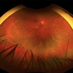
 Traction Detachment of Retina
Nov 14 2025 by Virginia Gebhart
50 year old female with proliferative diabetic retinopathy, large ridge of traction temporally, and significant band of fibrosis. Subretinal fluid throughout the macula. Due to traction, surgical repair not recommended at this time as it could worsen condition. Will observe closely. BCVA CF @ 1 ft.
Photographer: Virginia Gebhart, Retina Consultants of Carolina
Imaging device: Optos California
Condition/keywords: fibrosis, proliferative diabetic retinopathy (PDR), traction detachment, Traction retinal detachment
-
 Coats' Disease
Sep 2 2025 by Drew Mitchell
Optos color photograph of a young boy with Coats disease. Extensive subretinal exudation that is encroaching towards macula. There are peripheral berry aneurysms with localized area of subretinal fluid. Discussed treatment options including laser photocoagulation of aneurysms. Risks benefits and alternatives discussed including possible need for cryo.
Photographer: Drew Mitchell, OCT-C
Imaging device: Optos California
Condition/keywords: Coats' disease
-
 Small Retinoschisis
Aug 30 2025 by Gustavo Uriel Fonseca Aguirre
This longitudinal B-scan reveals a small inferotemporal peripheral retinoschisis, appearing as a smooth, thin-walled bipartite retinal separation without associated subretinal fluid or vitreous traction. The lesion demonstrates characteristic acoustic homogeneity and minimal mobility on dynamic evaluation.
Photographer: Gustavo U. Fonseca Aguirre, Hospital Conde de Valenciana, Ciudad de México
Condition/keywords: retinoschisis
-
 Amelanotic Melanoma
Aug 12 2025 by César Adrián Gómez Valdivia, MD
This FAF image reveals a hypoautofluorescent mass with areas of dense hyperautofluorescent stippling—a classic pattern suggestive of an amelanotic choroidal melanoma. Amelanotic melanoma is a rare variant of uveal melanoma, accounting for only a minority of cases. Unlike pigmented melanomas, these lesions lack melanin, making them more challenging to detect on conventional color fundus imaging. FAF Characteristics: • Central hypoautofluorescence: due to loss or compression of the RPE • Peripheral hyperautofluorescent speckling: consistent with lipofuscin accumulation or RPE disruption • Often associated with subretinal fluid or orange pigment seen clinically Location: Juxtapapillary, with potential optic nerve involvement—a factor that complicates both diagnosis and
Photographer: @eyemissu2
Imaging device: California ICG OPTOS
Condition/keywords: amelanotic melanoma
-
 Amelanotic Melanoma
Aug 12 2025 by César Adrián Gómez Valdivia, MD
This case highlights an amelanotic melanoma, an atypical presentation of a choroidal melanoma lacking the characteristic pigmentation. These lesions can easily be mistaken for choroidal hemangiomas, metastases, or inflammatory masses. Clinically, the lesion appears as a dome-shaped, yellowish subretinal mass, often associated with subretinal fluid, lipofuscin deposition, or retinal detachment. The absence of pigment can delay diagnosis, making multimodal imaging essential. Diagnostic tools: • B-scan ultrasound: low to medium internal reflectivity • OCT: overlying subretinal fluid and RPE elevation • FAF: orange pigment and RPE disruption • ICG/FA: variable, often hypofluorescent core Important: Prompt referral to ocular oncology is critical for management and prognosis.
Photographer: @eyemissu2
Imaging device: TOPCON TRC-50DX
Condition/keywords: amelanotic melanoma
-
 Chronic RD with Retinal Dialysis
Jul 23 2025 by Virginia Gebhart
64 year old female with chronic retinal detachment from head trauma 41 years ago. Peripheral scarring from 6:00 to 11:00 with area of subretinal fluid inferotemporally, well demarcated with subretinal bands. Retinal dialysis inferotemporal from 7:00 to 9:00. No surgical repair needed or recommended at this time.
Photographer: Virginia Gebhart, Retina Consultants of Carolina
Imaging device: Optos California
Condition/keywords: chronic retinal detachment, demarcation, RD, Retinal Detachment, retinal dialysis, subretinal bands
-
 Choroidal Melanoma (USG)
Jul 5 2025 by Gustavo Uriel Fonseca Aguirre
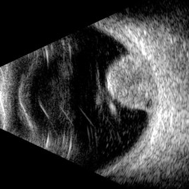
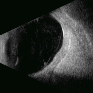
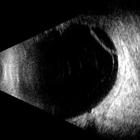
This B-mode transverse ultrasound scan reveals a mushroom-shaped choroidal tumor in the inferior nasal quadrant adjacent to the optic nerve head. The lesion appears solid with homogeneous internal reflectivity and is associated with minimal surrounding subretinal fluid and scleral excavation. It measures 6.54 mm in height × 7.52 mm in base diameter (transverse view) and extends 9.52 mm longitudinally. The vitreous contains abundant punctate opacities consistent with pigment dispersion. The retina and choroid remain attached elsewhere.
Photographer: Gustavo U. Fonseca Aguirre, Hospital Conde de Valenciana, Ciudad de México
Condition/keywords: choroidal melanoma
-
 Giant Retinal Tear
Jul 5 2025 by Gustavo Uriel Fonseca Aguirre
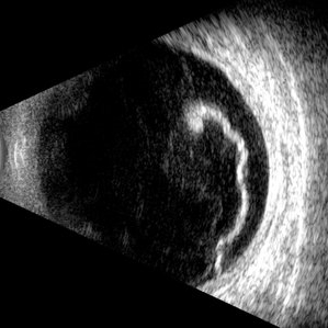
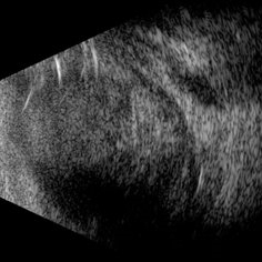
This B-mode longitudinal ultrasound scan reveals a giant retinal tear, demonstrating a circumferential retinal flap with rolled edges extending over M-X to M-I. The vitreous shows diffuse hemorrhage and anterior-posterior traction strands inserting at the tear margins. The remaining retina appears attached without subretinal fluid.
Photographer: Gustavo U. Fonseca Aguirre, Hospital Conde de Valenciana, Ciudad de México
Condition/keywords: giant retinal tear
-
 Retinal Dialysis
Jul 5 2025 by Gustavo Uriel Fonseca Aguirre
This B-mode longitudinal ultrasound scan demonstrates a retinal dialysis, appearing as a linear discontinuity at the ora serrata with associated vitreous base avulsion. The scan reveals mild subretinal fluid extending from the dialysis site with macular involvement.
Photographer: Gustavo U. Fonseca Aguirre, Hospital Conde de Valenciana, Ciudad de México
Condition/keywords: retinal dialysis
-
 Diabetic Macular Edema
Jul 3 2025 by Gustavo Uriel Fonseca Aguirre
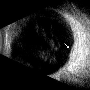
This B-mode longitudinal ultrasound scan demonstrates diabetic macular edema with mild subretinal fluid accumulation, appearing as a subtle hypoechoic space beneath the neurosensory retina. The macular region shows retinal thickening and heterogeneous medium reflectivity, consistent with active exudative changes (arrow). No vitreomacular traction is observed.
Photographer: Gustavo U. Fonseca Aguirre, Hospital Conde de Valenciana, Ciudad de México
Condition/keywords: diabetic macular edema
-
 Macular Retinoschisis
Jul 3 2025 by Gustavo Uriel Fonseca Aguirre
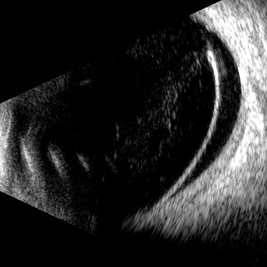
This B-mode longitudinal ultrasound scan reveals macular retinoschisis, demonstrating a characteristic splitting of retinal layers with a smooth, dome-shaped elevation. The lesion shows maintained structural integrity of both inner and outer retinal walls without associated subretinal fluid or vitreous traction.
Photographer: Gustavo U. Fonseca Aguirre, Hospital Conde de Valenciana, Ciudad de México
Condition/keywords: macular retinoschisis
-
 OCT Choroidal Rupture
Jun 26 2025 by Hector Gabriel Moreno Solano, MD, MHA
High-resolution OCT of the right eye shows a localized disruption of the retinal pigment epithelium (RPE)–Bruch’s membrane complex, consistent with a choroidal rupture. There is loss of the normal outer retinal architecture over the lesion, with focal elevation and irregularity of the underlying RPE. Hyperreflective material is noted at the level of the break, without associated subretinal fluid or signs of active choroidal neovascularization.
Photographer: Hector Gabriel Moreno Solano, Instituto Mexicano de Oftalmología “IMO I.A.P”
Imaging device: REVO
Condition/keywords: Choroidal Rupture, OCT
-
 Complex Retinal Detachment with PVR and Starfold
Jun 6 2025 by Jenn Geelan
57 year old male with a Complex Retinoschisis related retinal detachment with PVR and a Posterior Star Fold
Photographer: Jenn Geelan, Retina-Vitreous Surgeons of CNY
Imaging device: Optos California
Condition/keywords: proliferative vitreoretinopathy (PVR), rare, Retinal Detachment, retinoschisis, Starfolds, subretinal fluid
-
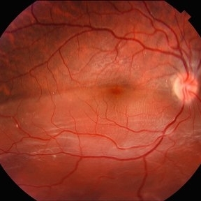
 Retinal Detachment
Jun 5 2025 by César Adrián Gómez Valdivia, MD
Fundus Photograph of a 19 year-old male patient with a RRD due to a Retinal Dialysis. Subretinal fluid and retinal folding can be appreciated.
Photographer: @eyemissu2
Imaging device: OPTOS
Condition/keywords: retinal detachment
-
 Retinal Detachment
Jun 5 2025 by César Adrián Gómez Valdivia, MD
Fundus Photograph of a 19 year-old male patient with a RRD due to a Retinal Dialysis. Subretinal fluid and retinal folding can be appreciated.
Photographer: @eyemissu2
Imaging device: TOPCON TRC-50DX
Condition/keywords: retinal detachment
-
 Scleral Rupture
May 9 2025 by Gustavo Uriel Fonseca Aguirre
This B-mode longitudinal ultrasound scan reveals dense vitreous hemorrhage, subretinal fluid, annular choroidal detachment, and scleral wall discontinuity with adjacent scleral folds. These findings indicate severe ocular trauma with probable scleral rupture and multi-compartment involvement.
Photographer: Gustavo U. Fonseca Aguirre, Hospital Conde de Valenciana, Ciudad de México
Condition/keywords: ocular trauma, scleral rupture
-
 Diabetic Macular Edema
Apr 28 2025 by Gustavo Uriel Fonseca Aguirre
This B-mode longitudinal ultrasound scan demonstrates irregular macular thickening with homogeneous medium-to-high internal reflectivity, consistent with diabetic macular edema. The lesion shows poorly defined borders and absence of cystic spaces or subretinal fluid on dynamic evaluation.
Photographer: Gustavo U. Fonseca Aguirre, Hospital Conde de Valenciana, Ciudad de México
Condition/keywords: diabetic macular edema
-
 Retinoschisis
Apr 21 2025 by Gustavo Uriel Fonseca Aguirre
This B-mode longitudinal ultrasound scan reveals a peripheral temporal retinoschisis, demonstrating a characteristic thin, dome-shaped separation of the retinal layers without associated subretinal fluid or vitreous traction. The lesion shows smooth, convex contours with maintained structural integrity of both retinal layers.
Photographer: Gustavo U. Fonseca Aguirre, Hospital Conde de Valenciana, Ciudad de México
Condition/keywords: retinoschisis
-
 Choroidal Osteoma
Apr 17 2025 by Gustavo Uriel Fonseca Aguirre
Scanning laser ophthalmoscopy reveals a well-circumscribed, yellowish-white choroidal osteoma overlying the macular region and extending into the inferior temporal vascular arcade. Retinal vessels course normally over the tumor surface, with no evidence of subretinal fluid or hemorrhage. The surrounding retina shows preserved architecture without secondary degenerative changes.
Photographer: Gustavo U. Fonseca Aguirre, Hospital Conde de Valenciana, Ciudad de México
Condition/keywords: choroidal osteoma, macular choroidal osteoma
-
 Treated Melanoma with Iluvien Implant
Apr 9 2025 by Virginia Gebhart
62 year old female 4 mo s/p brachytherapy for amelanotic choroidal melanoma. Iluvien implant given 4 wks s/p plaque removal, lesion is stable with resolved exudative detachment and subretinal fluid
Photographer: Virginia Gebhart, Retina Consultants of Carolina
Imaging device: Optos California
Condition/keywords: amelanotic melanoma, brachytherapy, choroidal melanoma, Iluvien, melanoma
-
 Chronic Central Serous Chorioretinopathy (CSCR)
Mar 31 2025 by Niloofar Piri, MD
Fundus Autofluorescence image of the right eye demonstrates classic guttering with hyper autofluorescence consistent with chronic CSCR. Guttering occurs where subretinal fluid migrates inferiorly due to gravity and stressed RPE cells accumulate lipofuscin material from high turnover of photoreceptor outer segments.
Photographer: Stefan Raev, COT; Saint Louis University
Condition/keywords: central serous chorioretinopathy (CSCR), Chronic CSR, Guttering
 Loading…