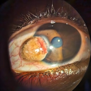
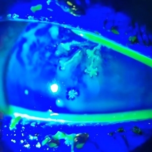
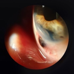
Search results (118 results)
-
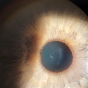
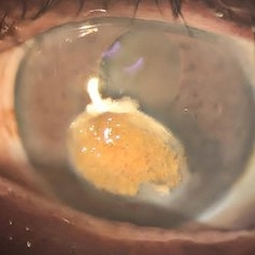
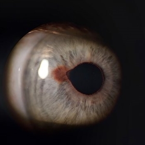
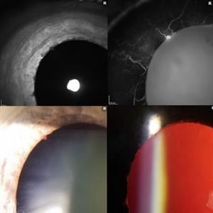
 Retisert Implant Migration Into the Anterior Chamber
Feb 11 2025 by Niloofar Piri, MD
Slit Lamp photograph demonstrating spontaneous dislocation and migration of old Retisert implant into the anterior chamber inferiorly with secondary corneal decompensation. Please notice that patient is aphakic. Implant was removed surgically.
Photographer: Hossein Asghari MD, Saint Louis University
Condition/keywords: Corticosteroid implant, implant migration, Retisert
-
 Iris Nevus
Jan 28 2025 by Korey Starkey
Slit-lamp image of an 89-year-old patient with an iris nevus. Nevus appeared stable on exam, will continue to monitor.
Photographer: Korey Starkey
Imaging device: Slit lamp camera
Condition/keywords: ectropion uveae, iris nevus, slit lamp photo
-
 Iris Melanoma
Jan 28 2025 by Korey Starkey
Slit-lamp image of 90-year-old patient with iris melanoma and new hemorrhage affecting the right eye. Patient re-presented after nearly 1 year, now seeking treatment. Given iris location of tumor, multiple clock hours of iris involved, and increase in size of the known malignant transformation; safest approach was enucleation.
Photographer: Korey Starkey
Imaging device: Slit lamp camera
Condition/keywords: anterior chamber, hemorrhage, iris melanoma, slit lamp photo
-
 Epicapsular Stars
Jan 28 2025 by Korey Starkey
Epicapsular stars and cataract noted in natural lens of 68-year-old patient.
Photographer: Korey Starkey
Imaging device: Slit lamp camera
Condition/keywords: cataract, chicken tracks, epicapsular stars, slit lamp photography
-
 Vitreous Prolapse
Jan 28 2025 by Korey Starkey
Slit lamp image of a 62-year-old patient presented at first visit with vitreous prolapse due to mechanical complications from IOL placement. IOP was being managed with drops, vision was 20/20, patient opted for surgery due to constant haze in vision.
Photographer: Korey Starkey
Imaging device: Slit lamp camera
Condition/keywords: slit lamp photo, vitreous prolapse
-
 PFO Bubbles
Oct 24 2024 by Korey Starkey
Slit lamp photograph of a 23 year old female with PFO bubbles inferiorly in the AC. Discussed surgical intervention to remove PFO from AC and vitreous cavity in future.
Photographer: Korey Starkey
Imaging device: Slit lamp camera
Condition/keywords: anterior chamber, PFO, slit lamp photography, submacular perfluorocarbon liquid (PFO)
-
 Kayser-Fleischer Ring
Oct 5 2024 by DR Rohit Gupta
Slit lamp photograph of 12 year old male patient suffering from Wilson's disease. This patient presented in opd with extended phalanges, pain in abdomen and disoriented.
Photographer: Dr Rohit gupta
Imaging device: Samsung S21
Condition/keywords: Kayser-Fleischer ring, Kf ring
-
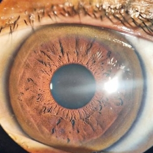
 Radial Keratotomy
Sep 28 2024 by DR Rohit Gupta
A slit lamp photograph of 46 year-old female patient operated for high hypermetropia 10 years back . On slit lamp examination hexagonal pattern of radial incisions can be seen.
Photographer: Dr Rohit gupta
Condition/keywords: hypermetropia, hyperopia, Radial keratotomy, refractive surgery
-
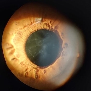
 Spheroidal Degeneration
Sep 28 2024 by DR Rohit Gupta
Slit lamp photograph of a 68 year-old male patient presented with diminution of vision and foreign body sensation. On examination brown cataract with yellowish globular degeneration seen on cornea.
Photographer: Dr Rohit gupta
Imaging device: Samsung S21
Condition/keywords: Spheroidal degeneration
-
 Limbal Dermoid
Sep 25 2024 by DR Rohit Gupta
Slit lamp photograph of a 22 year-old female presenting with a swelling over cornea which on examination appears be to be Limbal dermoid.
Photographer: Dr Rohit gupta
Imaging device: Samsung S21
Condition/keywords: dermoid, limbus, lipodermoid
-
 Herpes Simplex
Sep 24 2024 by DR Rohit Gupta
Slit lamp photograph of a 32 year-old male presented with redness, photophobia, and pain in left eye.
Photographer: Dr Rohit gupta
Imaging device: Samsung S21
Condition/keywords: corneal ulcer, Herpes, herpes dendrite, Herpes simplex infection
-
 Herpetic Corneal Ulcer
Sep 24 2024 by DR Rohit Gupta
Slit lamp photograph of 32 year old male presented with herpetic corneal ulcer on staining with fluorescein dye under cobalt blue filted dendrits can be seen.
Photographer: Dr Rohit gupta
Imaging device: Samsung S21
Condition/keywords: corneal ulcer, dendritic keratitis, herpes dendrite, Herpes simplex infection, Herpes zoster, staining
-
 Scleral Ectasia Post Radiation for Iris Melanoma
Jul 5 2024 by Zach Seim
Slit-Lamp Photograph of a 52 year old male with Scleral Ectasia post radiation for Iris Melanoma.
Photographer: Zach Seim
Imaging device: Slit Lamp Photography on Samsung Galaxy 7
Condition/keywords: Iris, iris melanoma, scleral ectasia, slit lamp photo, slit lamp photography
-
 Congenital Nuclear Cataract
Jul 5 2024 by Zach Seim
This is a slit-lamp photograph of a 10 year old female with a congenital nuclear cataract OD. Patient presented with VA Dsc 20/200. Patient was counseled on surgical options.
Photographer: Zach Seim
Imaging device: Slit Lamp Photography on Samsung Galaxy 7
Condition/keywords: cataract, congenital cataract, nuclear sclerosis, right eye, slit lamp photo
-
 Iris Nevus
Jul 3 2024 by Zach Seim
Slit Lamp Photograph of an 88 year old man with an Iris Nevus. Patient presented with DCC 20/60+1. Plan to monitor.
Photographer: Zach Seim
Imaging device: Slit Lamp photography with Samsung Galaxy 7
Condition/keywords: iris, iris nevus, nevus, right eye, slit lamp photo, slit lamp photography
-
 Dislocated IOL
Jun 4 2024 by Marlee Curnutt
Slit lamp photo of a 64 year old woman presenting with worsening vision and depth perception after trauma induced by a dog, which dislocated her IOL. The patient's IOL haptic was seen in the AC, and almost abutting cornea. Patient's vision upon presentation was DCC CF@1 feet. Patient was counseled and underwent an IOL exchange.
Photographer: Marlee Curnutt, COA
Imaging device: Galaxy A42
Condition/keywords: dislocated intraocular lens (IOL), haptic, IOL, right eye, slit lamp photo, slit lamp photography, trauma
-
 Asteroid Hyalosis
Apr 9 2024 by Hector Gabriel Moreno Solano, MD, MHA
Slit lamp photograph of a 48-year-old female patient with long-standing diabetes attending consultation due to the sensation of moving spots in her vision.
Photographer: Héctor Gabriel Moreno-Solano
Condition/keywords: asteroid hyalosis, diabetes, slit lamp photo
-
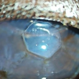
 Perfluorocarbon Bubbles in Anterior Chamber
Mar 23 2024 by Victoria Fernanda Coba Caizaluisa
Slit lamp photography: bubbles of PFCL in the anterior chamber after one month of retinal detachment surgery.
Photographer: Victoria Coba, University of Buenos Aires, Centro de Ojos Lanus
Condition/keywords: anterior chamber, perfluorocarbon fluid, retained perfluorocarbon, Retinal Detachment
-
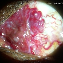 Conjunctival AV Malformation
Dec 18 2023 by siddharth sheth
33 year old male presented with a complaint of redness since 15 years in left eye.
Photographer: Gaurav Kamble, Isha Netralaya
Imaging device: Dyanmic slit lamp imaging
Condition/keywords: conjunctival AV malformation, slit lamp photo, slit lamp photography, unilateral
-
 Conjunctival involvement uveal lymphoma
Jan 20 2023 by Elaine Michele Binkley, MD
Slit lamp photograph shows characteristic "salmon-patch" conjunctival lesion in the setting of uveal marginal zone lymphoma with conjunctival involvement.
Photographer: Brice Critser, University of Iowa
Condition/keywords: Uveal Lymphoma
-
 Iris Vascular Tuft
Jul 5 2022 by Olivia Rainey
Anterior segment imaging of a 66-year-old male with Vascular Disorders of Iris and Ciliary Body affecting his right eye. The physician stated that the findings are most consistent with a benign vascular tuft at the pupillary margin. The patient presented at the office with 20/20 vision in both eyes and had no ocular complaints at the time of his appointment.
Photographer: Olivia Rainey, OCT-C, COA
Imaging device: Heidelberg Spectralis, Slit Lamp with Samsung Galaxy 7
Condition/keywords: anterior segment, fluorescein angiogram (FA), heidelberg spectralis, infrared image, near infrared autofluorescence (NIRAF), slit lamp photo, vascular anomaly, vascular disorders of iris and ciliary body, vascular tuft
-
 Koeppe Iris nodules
Apr 14 2022 by Divya Jain
Slit Lamp photograph of a 33 year old woman with first episode of acute granulomatous anterior uveitis showing mutton fat KP'S and Koeppe's nodules.
Photographer: Divya Jain
Condition/keywords: acute anterior uveitis
-
 Acute Anterior Uveitis
Apr 14 2022 by Divya Jain
Anterior Segment Slit Lamp photograph of a 33 year old woman with first episode of acute granulomatous anterior uveitis showing circumcorneal congestion, mutton fat KP'S, 3+ cells, 2+ flare and Koeppe's nodules at pupillary margin.
Photographer: Divya Jain
Condition/keywords: acute anterior uveitis
-
 VZV Keratitis: Slit lamp Photo
Dec 8 2021 by Wen Hu
Slit lamp photograph of a 29-year-old man with VZV keratitis, who later developed choroidopathy.
Condition/keywords: varicella zoster virus (VZV), Varicella Zoster Virus Keratitis
-
 Intravitreal Anti -VEGF Complication: Posterior Capsular Breach
Dec 6 2021 by Nizamuddin HM Shaik, MD, FRCS
Slit lamp photo of patient with posterior capsular breach following an intravitreal anti-VEGF injection.
Photographer: Nizamuddin HM Shaik, MD
Condition/keywords: Intravitreal Anti VEGF Complication, posterior capsular breach
 Loading…