Search results (76 results)
-
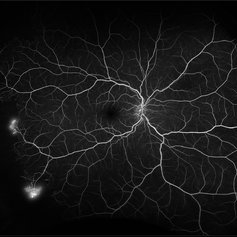
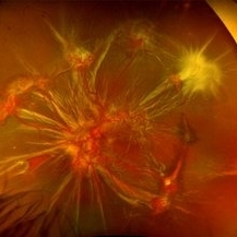
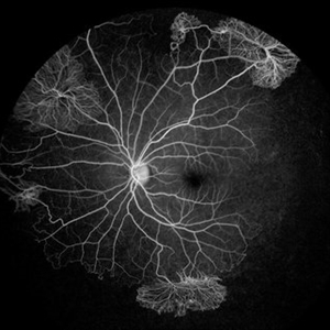 Proliferative Sickle Cell Retinopathy
Jul 8 2025 by Niloofar Piri, MD
Mid AV phase fluorescein angiogram of a 13 yo AA male with SC disease demonstrating multiple classic sea fan neovascularization with peripheral capillary non perfusion (CNP). CNP is more obvious in this image involving the temporal retina and inferonasal retina.
Photographer: Stefan Raev, COT, Saint Louis University
Condition/keywords: Proliferative sickle cell retinopathy, proliferative sickle retinopathy, sickle cell retinopathy
-
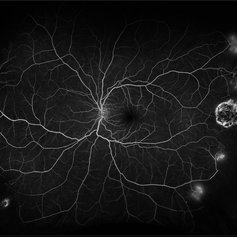
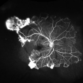 Proliferative Sickle Retinopathy
Jun 13 2025 by Brandon I Fram, MD
30 year-old with HbSC sickle retinopathy found to have profound retinal ischemia and florid peripheral neovascularization.
Imaging device: Fluorescein Angiography
Condition/keywords: proliferative sickle retinopathy, retinal ischemia, sea fan, sickle cell retinopathy
-
 Sickle Cell Retinopathy
Feb 24 2025 by Kimberly Wakester
Optomap RGB image of an 24-year-old woman with sickle cell retinopathy in both eyes. There is overall progression of the ischemic vessels and vascular drops out compared to previous images completed in 2021. Oral FA was completed and shows possible progression of peripheral non-perfusion but difficult to determine due to drinking FA dye and images not being as bright. On Clinical exam there is no evidence of NV, RD, or RT in either eye. Patient understands the need for continued follow up care and the likely need for PRP laser in both eyes.
Photographer: Kimberly Wakester, COA
Imaging device: Optos California
Condition/keywords: sickle cell retinopathy
-
 Proliferative Sickle Cell Retinopathy
Jan 27 2025 by Virginia Gebhart
61 year-old with proliferative sickle cell retinopathy s/p cryotherapy to peripheral fibrotic NV. Eye is stable with resolving exudates and maturing cryo scar. BCVA 20/40
Photographer: Virginia Gebhart, Retina Consultants of Carolina
Imaging device: Optos California
Condition/keywords: cryotherapy, fibrotic neovascularization, sickle cell retinopathy
-
 Sickle-Cell Retinopathy
Jan 22 2025 by Virginia Gebhart
Fluorescein angiogram of 54 year old female with non-diabetic proliferative retinopathy. Recent labs confirm sickle-cell disease. FA shows temporal peripheral non perfusion with NV. S/p PRP with retrobulbar block
Photographer: Virginia Gebhart, Retina Consultants of Carolina
Imaging device: Optos California
Condition/keywords: FA, Fluorescein angiography, Neovascularisation elsewhere (NVE), non-perfusion, Nose, pan-retinal photocoagulation (PRP), PRP, sickle cell retinopathy
-
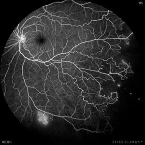 Proliferative Sickle Cell Retinopathy
Nov 4 2024 by Ricardo Leitão Guerra
Peripheral non perfusion and sea-fan in a case of sickle cell retinopathy.
Photographer: Ricardo Leitão Guerra, Leitão Guerra - Oftalmologia, Salvador-Brazil
Imaging device: Clarus 700 - Zeiss
Condition/keywords: Fluorescein angiography, Sickle cell Retinopathy
-
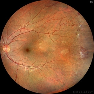 Sickle Cell Retinopathy (Proliferative) - Color
Oct 27 2023 by Ricardo Leitão Guerra
Proliferative sickle cell retinopathy.
Photographer: Ricardo Luz Leitão Guerra, Leitão Guerra - Oftalmologia, Salvador - Brazil
Imaging device: Clarus 700
Condition/keywords: Iridescent spots, non-perfusion, sea fan, sickle cell retinopathy
-
 Sickle cell retinopathy (Proliferative) - FA
Oct 27 2023 by Ricardo Leitão Guerra
Fluorescein angiography in a case of proliferative sickle cell retinopathy.
Photographer: Ricardo Luz Leitão Guerra, Leitão Guerra - Oftalmologia, Salvador - Brazil
Imaging device: Clarus 700
Condition/keywords: Iridescent spots, non-perfusion, sea fan, sickle cell retinopathy
-
 Sickle Cell Hemoglobinopathy
Aug 21 2023 by Harsh Vardhan Singh, MS
A 12-year-old male with a known case of sickle cell hemoglobinopathy with symmetrical dark without pressure in mid-periphery in both eyes
Photographer: Dr Harsh Vardhan Singh
Condition/keywords: Dark without Pressure, Sickle cell Retinopathy
-
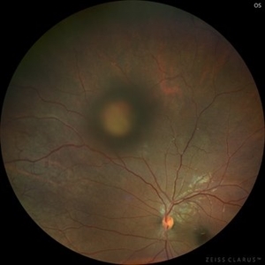 Salmon Patch
Jul 25 2023 by Kamal Kishore, MD, MBBS
A 14-year-old male with proliferative sickle cell retinopathy (PSR) with a salmon patch lesion at the superonasal periphery
Photographer: Kim Grabill, COA, Illinois Retinal and Eye Associates, Peoria, IL, USA
Imaging device: Zeiss Clarus
Condition/keywords: salmon patch, sickle cell retinopathy
-
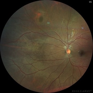 Black Sunburst in Proliferative Sickle Cell Retinopathy
Jul 25 2023 by Kamal Kishore, MD, MBBS
A 17-year-old male with a black sunburst lesion at superonasal periphery.
Photographer: Jessi Wright
Imaging device: Zeiss Clarus
Condition/keywords: Black Sunburst, sickle cell retinopathy
-
 Proliferative Sickle Cell Retinopathy
Feb 1 2023 by Olivia Rainey
Ultra-widefield fluorescein angiography of a 25-year old male with Proliferative Sickle Cell Retinopathy affecting his right eye. Patient stated that he was born with Sickle disease (SC), and has yearly eye exams. He noted no vision concerns over the last year but has typically experienced sickle attacks about 1-2 per year. The physician noted that the fluorescein obtained showed peripheral nonperfusion affecting the patient's nasal and temporal retina as well as neovascularization affecting his left eye more than his right. He recommended pan retinal photocoagulation in his left eye for his temporal and nasal retina, as as well as his right eye following.
Photographer: Olivia Rainey, OCT-C, COA
Imaging device: Optos California
Condition/keywords: early phase, fluorescein angiogram (FA), fluorescein leakage, neovascularization (NV), non-perfusion, proliferative retinopathy, right eye, sickle cell retinopathy, ultra-wide field imaging, ultra-widefield image
-
 Proliferative Sickle Cell Retinopathy
Feb 1 2023 by Olivia Rainey
Ultra-widefield fluorescein angiography of a 25-year old male with Proliferative Sickle Cell Retinopathy affecting his left eye. Patient stated that he was born with Sickle disease (SC), and has yearly eye exams. He noted no vision concerns over the last year but has typically experienced sickle attacks about 1-2 per year. The physician noted that the fluorescein obtained showed peripheral nonperfusion affecting the patient's nasal and temporal retina as well as neovascularization affecting his left eye more than his right. He recommended pan retinal photocoagulation in his left eye for his temporal and nasal retina, as as well as his right eye following.
Photographer: Olivia Rainey, OCT-C, COA
Imaging device: Optos California
Condition/keywords: early phase, fluorescein angiogram (FA), fluorescein leakage, left eye, neovascularization (NV), proliferative retinopathy, sickle cell retinopathy, ultra-wide field imaging, ultra-widefield image
-
 Sickle Cell Retinopathy - Optos Wide-field
Nov 14 2022 by Bryon R McKay, MD, PhD, FRCSC, DRCPSC - Retina
28 year-old female with floaters, History of Sickle Cell SC disease. No previous retinal treatments
Photographer: Dr. Bryon McKay
Imaging device: Optos California
Condition/keywords: sickle cell retinopathy
-
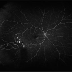
 Sickle Cell Retinopathy
Nov 14 2022 by Bryon R McKay, MD, PhD, FRCSC, DRCPSC - Retina
28 year-old female with floaters, History of Sickle Cell SC disease. No previous retinal treatments - fluorescein angiography showing sea-fan neovascularization
Photographer: Dr. Bryon McKay
Imaging device: Optos California
Condition/keywords: sickle cell retinopathy
-
 Sickle Cell Retinopathy
Nov 14 2022 by Bryon R McKay, MD, PhD, FRCSC, DRCPSC - Retina
28 year-old female with floaters, History of Sickle Cell SC disease. No previous retinal treatments - Late angiogram with extensive leakage
Photographer: Dr. Bryon McKay
Imaging device: Optos California
Condition/keywords: sickle cell retinopathy
-
 Sickle Cell Retinopathy
Nov 5 2022 by Mateus Queiroz Corrêa, MD
19 -year-old young man with combined rhegmatogenous and tractional retinal detachment secondary to a proliferative sickle retinopathy ( stage V)
Photographer: Mateus Corrêa, Sorocaba Eye Bank Hospital
Imaging device: Optos California
Condition/keywords: Retinal detachment, sickle cell retinopathy
-
 Displaced & folded macula
Oct 10 2022 by Ricardo Leitão Guerra
Tractional retinal detachment due to sickle cell retinopathy leading to a displaced and folded appearance of the macula in this 36-yo male. Subretinal bands are also noticed crossing the macula towards inferior retinal detachment area.
Photographer: Ricardo Leitão Guerra
Imaging device: Clarus 700 - Zeiss
Condition/keywords: folds, sickle cell retinopathy, subretinal bands, tractional retinal detachment
-
 Proliferative Diabetic Retinopathy and SC Disease
Aug 27 2021 by Caesar K. Luo, MD, FASRS
53 year-old male with SC disease complicated by proliferative diabetic retinopathy with severe peripheral non perfusion and small, central retained island.
Photographer: Fred Hanamoto, Bay Area Retina Associates
Imaging device: Optos California
Condition/keywords: capillary nonperfusion, peripheral ischemia, proliferative diabetic retinopathy (PDR), retinal ischemia, sickle cell retinopathy
-
 Proliferative Diabetic Retinopathy and SC Disease
Aug 27 2021 by Caesar K. Luo, MD, FASRS
53 year-old male with SC disease complicated by proliferative diabetic retinopathy with severe peripheral non perfusion and vascular sclerosis.
Photographer: Fred Hanamoto, Bay Area Retina Associates
Imaging device: Optos California
Condition/keywords: ischemia, peripheral ischemia, proliferative diabetic retinopathy (PDR), sickle cell retinopathy
-
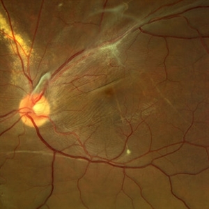
 Sickle Cell
Jul 6 2021 by Kristen Wagner
Fundus photo on the Clarus who is diagnosed with sickle cell retinopathy and has had laser treatment.
Photographer: Kristen Wagner COT, Tennessee Retina, Nashville TN
Condition/keywords: sickle cell retinopathy
-
 Retinal Hemorrhages
Mar 10 2021 by Kachelle Brown
Ultra widefield Fluorescein Angiography of a 48-year-old female with retinal hemorrhages affecting her right eye. Physician suspect sickle cell due to family history, and has ordered labs to rule out.
Photographer: Kachelle Brown
Imaging device: Optos California
Condition/keywords: fluorescein angiogram (FA), fluorescein leakage, Optos, retinal hemorrhage, sickle cell retinopathy, ultra-wide field imaging
 Loading…